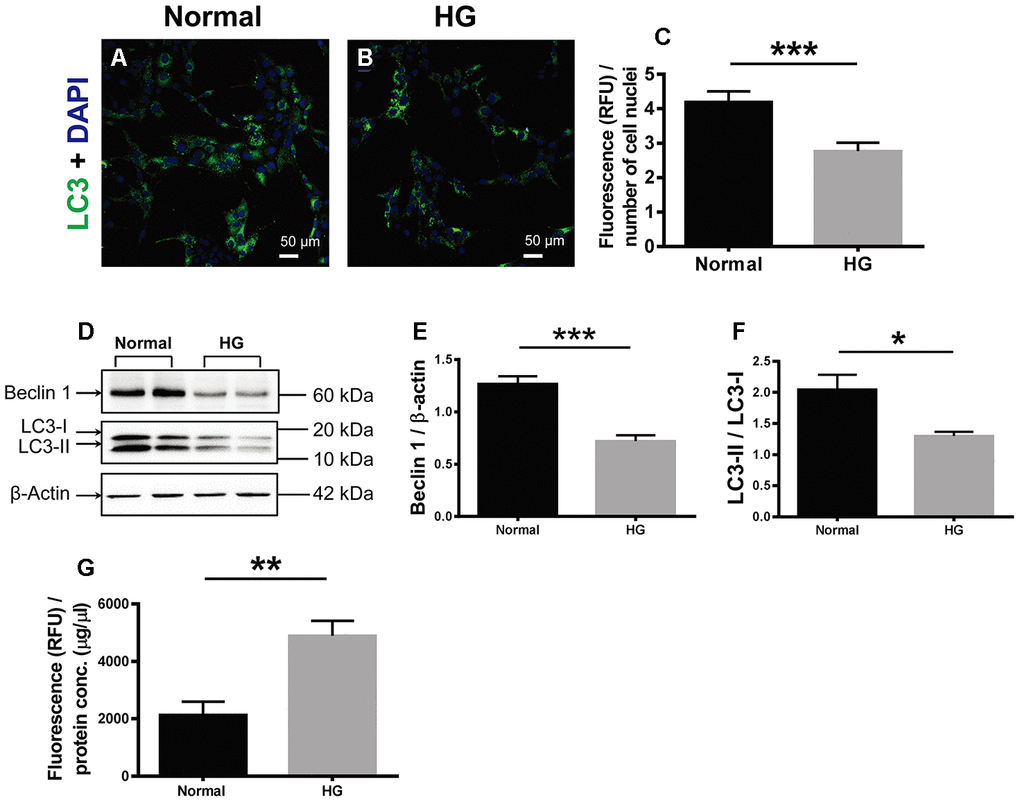
Figure 6.The effects of high glucose on autophagy and apoptosis of retinal vascular endothelial cells. (A, B) Immunofluorescence of LC3, an autophagosomal marker, in HG-treated RF/6A cells and normal controls. Scale bar = 50 μm (C) The fluorescence intensity of LC3 was quantified and normalized to the number of cell nuclei. (D) Western blot representatives of Beclin1, LC3-I, LC3-II, and β-actin. (E) Quantification of Beclin 1 protein levels in the RF/6A cells treated with HG-containing or normal culture media. (F) Ratio of LC3-II over LC3-I in the HG-treated RF/6A cells and normal controls. (G) The Caspase-3/7 activity (RFU) in the retinal vascular endothelial cells was quantified and normalized to the total protein concentration (μg/μl). n = 6 / group. Data are expressed as mean ± SEM. * P < 0.05, ** P < 0.01, *** P < 0.001.