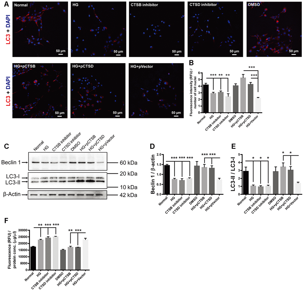
Figure 7.The effects of high glucose on autophagy and apoptosis were mimicked by CTSB and CTSD inhibitors and abrogated by their overexpression. (A) Immunofluorescence staining of LC3 in experimental groups. Scale bar = 50 μm (B) The fluorescence intensity of LC3 was quantified and normalized to the number of cell nuclei. (C) The representative pictures of Western blots of Beclin1, LC3-I, LC3-II, and β-actin. (D, E) Quantification of Beclin 1 protein levels and the ratio of LC3-II over LC3-I in the HG-treated RF/6A cells and normal controls. (F) The Caspase-3/7 activity (RFU) in the retinal vascular endothelial cells was quantified and normalized to the total protein concentration (μg/μl). n = 6 / group. Data are expressed as mean ± SEM. * P < 0.05, ** P < 0.01, *** P < 0.001.