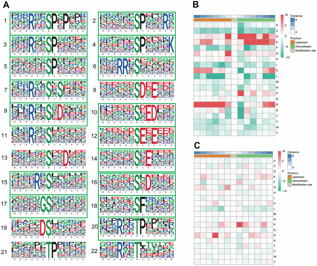
Figure 4.Motif analysis of the phosphosites. (A) Significantly enriched phosphorylation motifs extracted from the overrepresented phosphopeptide dataset by Motif-X. (1) -(19) Motifs from phosphoserine; (20) -(22) Motifs from phosphothreonine. Among these motifs, 6 are identified as 5 known phosphorylation motifs and 17 are newly identified (enclosed into green boxes). The detailed information and putative associated kinases are shown in Table 3. Motif enrichment heat map of phosphoserine (B) and phosphothreonine (C) upstream and downstream of all identified phosphorylation modification sites. Red indicates significant enrichment of the amino acid near the modification site, while green indicates significant reduction of the amino acid near the modification site.