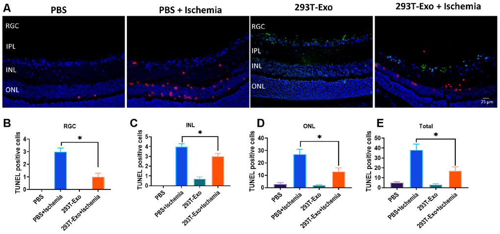
Figure 8.293T-Exo inhibited OGD-induced apoptosis in the retinae >in vivo (A) 293T-Exo inhibited OGD-induced apoptosis in the retinae, as measured by the TUNEL fluorescence assay. The nuclei were stained with DAPI (blue), the TUNEL-positive cells were stained with TUNEL dye (red), and 293T-Exo was stained with fluorescent-tag (green). Scale bar = 25 μm. (B–D) Number of TUNEL-positive cells in different structural layers in the retinae in response to treatment of PBS, PBS+ Ischemia, 293T-Exo, and 293T-Exo+ Ischemia, respectively. (E) Total number of TUNEL-positive cells in the retinae in response to treatment of PBS, PBS+ Ischemia, 293T-Exo, and 293T-Exo+ Ischemia. RGC: retinal ganglion cell; IPL: inner plexiform layer; INL: inner nuclear layers; ONL: outer nuclear layers. Data are presented as mean ± SD. *P < 0.05.