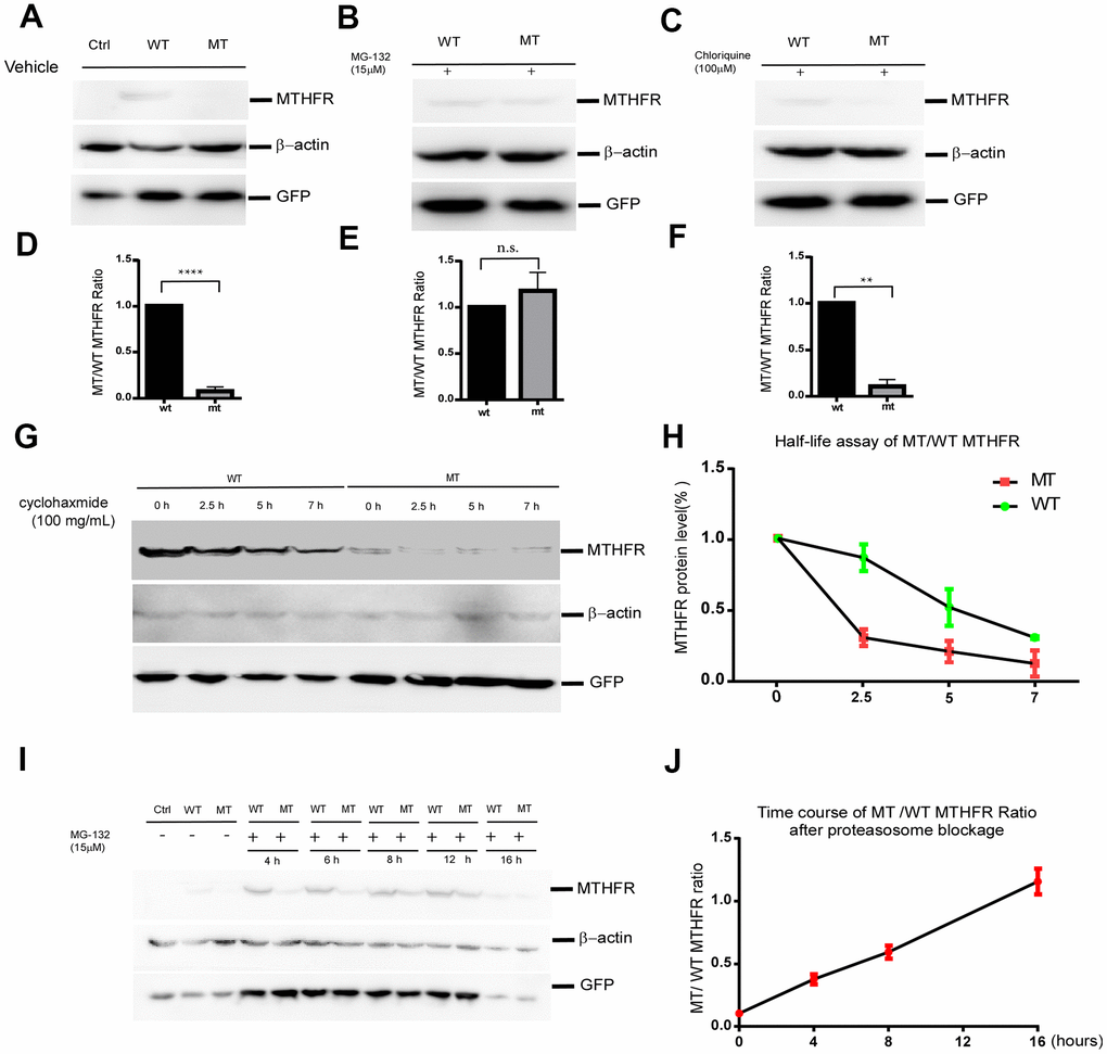
Figure 3.The p.Leu439Pro mutation in MTHFR facilitated MTHFR degradation via the proteasomal pathway. Western blot bands (A) and the quantification (D) demonstrated that at equal transfection efficiency (as indicated by GFP expression), the protein level of over-expressed MTHFRL439C was significantly lower than that of MTHFRWT (p<0.0001, n=3 replicates).Lysosome blockage by chloroquine failed to elevate the mutant MTHFR protein level (C, F. p=0.1746), whereas proteasome blockage by MG132 significantly increased MTHFRL439P, and abolished the difference between the wild type and mutant MTHFR (B, E. p<0.01, n=3 replicates). (G,H) Half-life assay of the WT and MTHFRL439P by using cycloheximide to block protein synthesis and chase the remaining protein level by western blot at 0, 2.5, 5, 7 hours later(G). The half-life of WT MTHFR is around 5 hours whereas the MTHFRL439P was almost depleted within 2.5 hours (H, n=3). (I, J) Representative Western blot result of WT and the mutant MTHFR protein expression on a time course of MG132 treatment(I), and quantifications (J), (n=2 replicates). All data are expressed as Mean±SEM. ****p<0.0001, **p<0.01 (by Student's t-test).