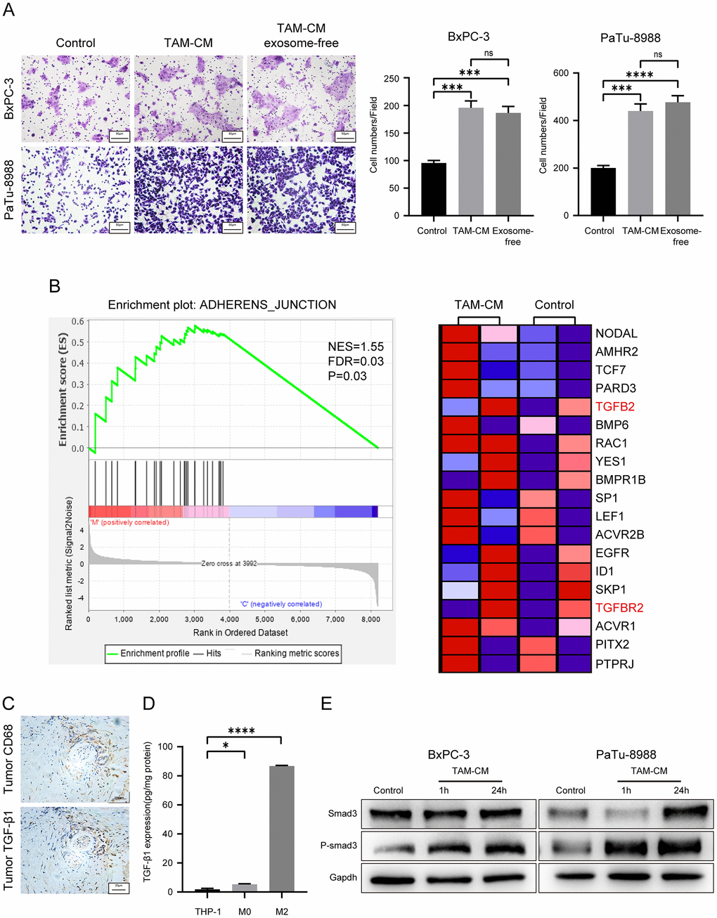
Figure 4.TAM-derived cytokines activate TGF-β signaling pathway in the PDAC cell lines. (A) Transwell assay results show the migration and invasiveness of BxPC-3 and PaTu-8988 cells incubated with exosome-free TAM-CM or normal TAM-CM. Scale bar = 50 μm; ***P < 0.001; ****P < 0.0001. (B) The enrichment plot shows significantly differentially expressed genes in PDAC cells treated with TAM-CM according to the Gene Set Enrichment Analysis (GSEA). (C) Representative images show IHC analysis of CD68 and TGF-β staining in the PDAC tissue sections. Scale bar = 20 μm. (D) ELISA analysis shows the levels of TGF-β secreted by THP-1, M0-type macrophages and M2-type macrophages. *P < 0.05; ****P < 0.0001. (E) Western blot analysis shows the levels of Smad3 and phospho-Smad3 proteins in control and TAM-CM treated BxPC-3 and PaTu-8988 cells.