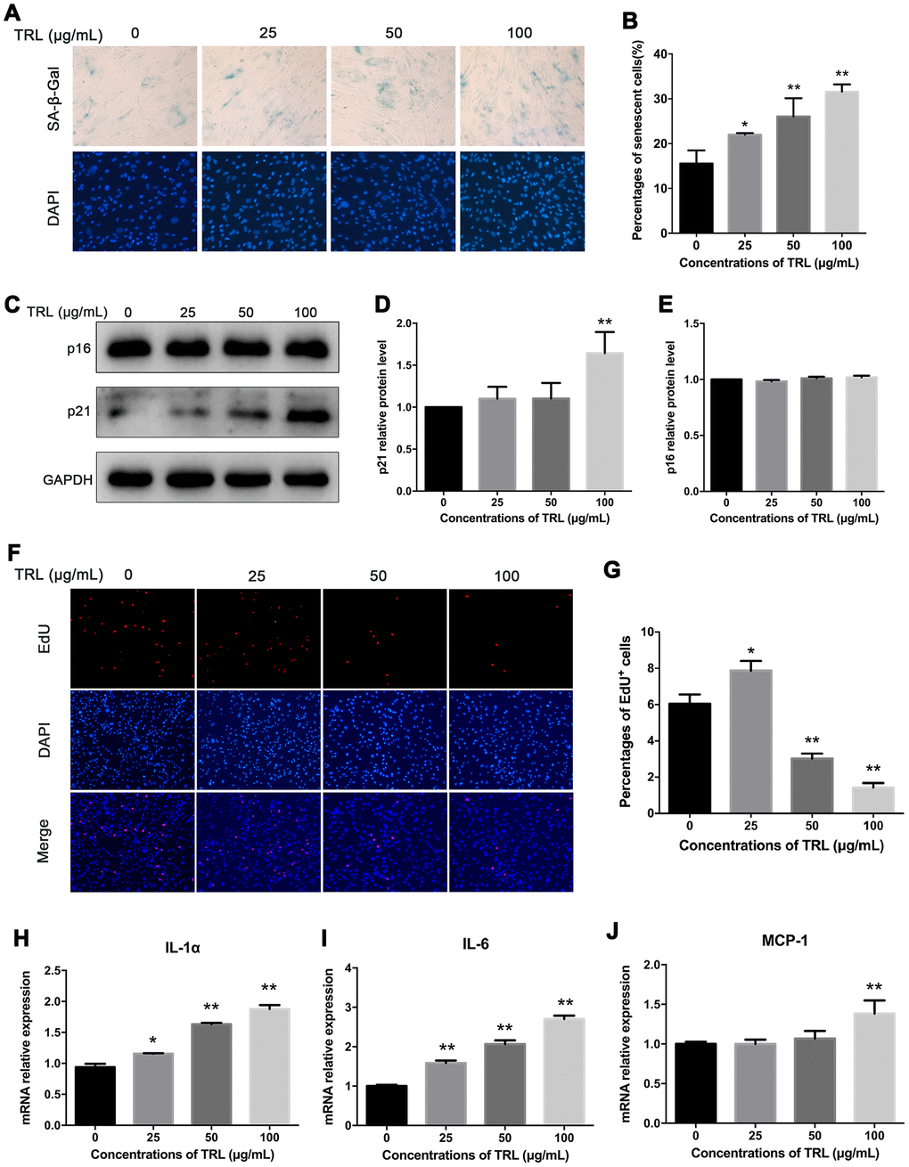
Figure 2.Postprandial TRL induced premature senescence and SASP in AMSCs. (A, B) AMSCs reached approximately 30%-40% culture-confluence were incubated with 0, 25, 50, or 100 μg/mL postprandial TRL for 8 d, and then SA-β-Gal (upper row) and DAPI (lower row) double staining was performed to detect the senescent cells and nuclei, respectively (A). Images were obtained under a microscope (×200 magnification). SA-β-Gal positive cells were counted manually by scanning a total of 200 cells in each sample (B). (C–E) Protein levels of senescent markers, p21 and p16, were detected using western blotting (C), and then the relative protein levels of p21 (D) and p16 (E) were analyzed using ImageJ. (F, G) The proliferation capacity of AMSCs incubated with different concentrations of postprandial TRL was measured using an EdU incorporation assay (F) and the EdU positive cells were counted using ImageJ (G). Images were obtained under a microscope (×100 magnification). (H–J) Expression levels of genes encoding senescence-related inflammatory cytokines, including IL-1α (H), IL-6 (I), and MCP-1 (J), were detected using qRT-PCR in AMSCs incubated with postprandial TRL at 0, 25, 50, or 100 μg/mL for 8 d. Data are expressed as mean ± SD (n ≥ 3). *P < 0.05, **P < 0.01 when compared with the control group.