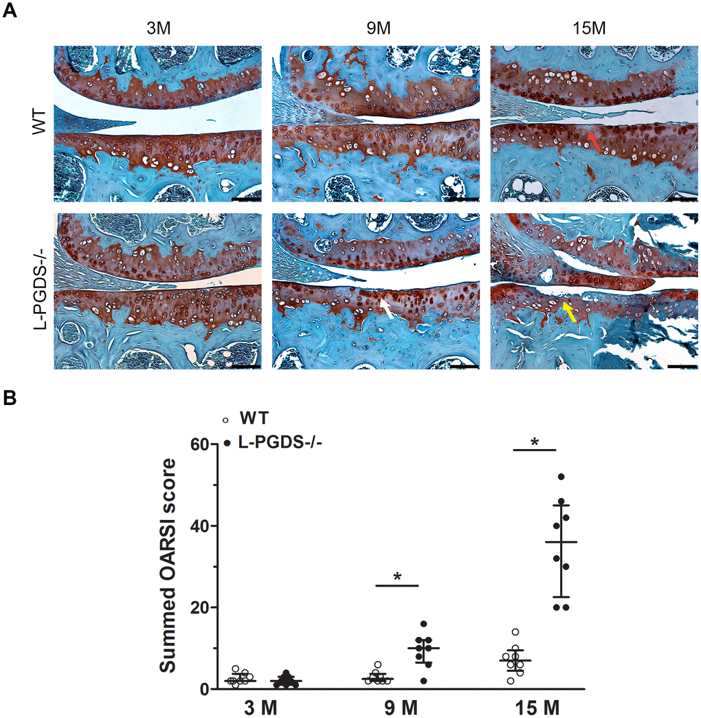
Figure 1.Deletion of L-PGDS accelerated cartilage erosion with age. (A) Coronal sections of whole knee joints from WT and L-PGDS-/- mice at ages 3, 9 and 15 months (n=8 mice/genotype/time point) were prepared and stained with Safranin O–fast green to assess the integrity of articular cartilage. The representative sections were selected based on the average score from each experimental group. Red arrow indicates loss of Safranin O staining. White arrow indicates areas of fibrillation and clefts. Yellow arrow indicates cartilage erosion. Scale bars=100 μM. (B) Summed histologic scores of knee cartilage from WT (open symbols) and LPGDS-/- (filled symbols) mice as determined using the OARSI scoring system. Results are presented as median with interquartile range. *P<0.05 versus WT mice.