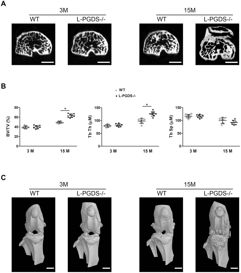
Figure 4.Micro-CT analysis of the subchondral bone of the tibial plateau of WT and L-PGDS-/- mice at 3 and 15 months. Knee joints from 3- and 15-month-old WT and L-PGDS-/- mice (n=8 mice/genotype/time point) were evaluated by micro-CT. (A) Representative axial micro-CT images of the subchondral bone compartment. Scale bars=1 mm. (B) Quantification of BV/TV, Tb.Th, and Tb.Sp in the subchondral bone region of the medial tibial plateau of WT (open symbols) and L-PGDS-/- (filled symbols) mice. Data are presented as mean ± SD. *p<0.05 versus WT mice. (C) Representative 3D reconstructions of the knee joints of WT and L-PGDS-/- mice at ages 3 months and 15 months PGDS-/- mice. Scale bars=1 mm.