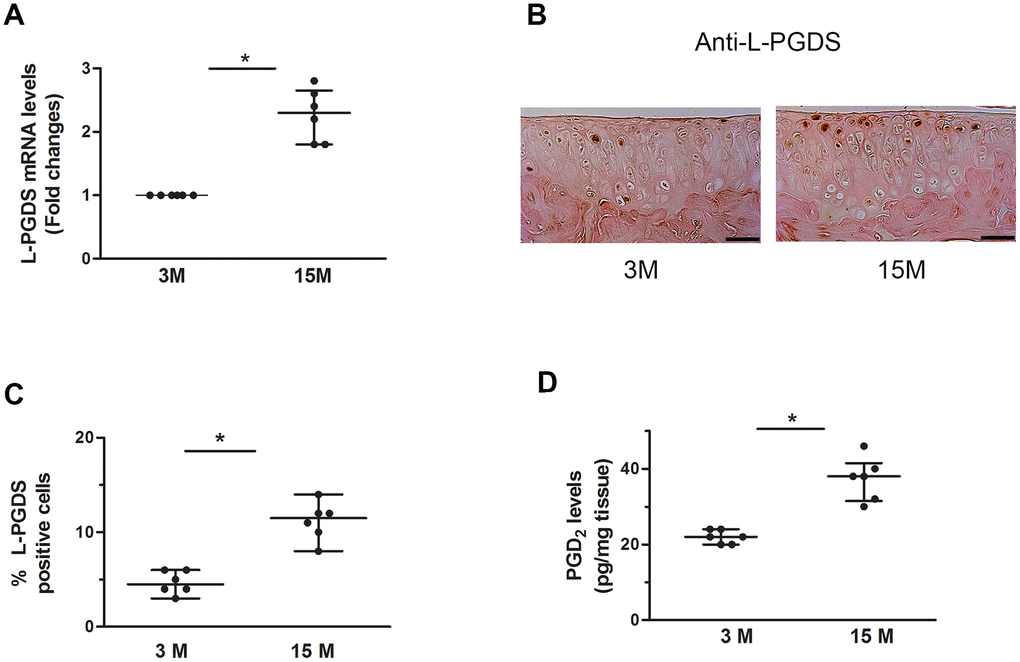
Figure 7.Increased expression of L-PGDS in cartilage of aged WT mice. (A) Total RNA was extracted from the joints of 3- and 15-month-old mice (n=6 mice/genotype/time point), and the levels of L-PGDS mRNA were determined by real-time RT-PCR. Results are expressed as -fold change, considering the value for 3-month-old mice as 1. (B) Representative images of immunohistochemical staining for L-PGDS in knee joints from of 3- and 15-month-old mice. Scale bars=100 μm. (C) Percentage of chondrocytes expressing L-PGDS in cartilage (n=6 mice/genotype/time point). Results are shown as median with interquartile range. (D) PGD2 levels in knee joint of 3- and 15-month-old mice (n=6 mice/genotype/time point), as determined by ELISA. *p<0.05 versus 3-month-old mice.