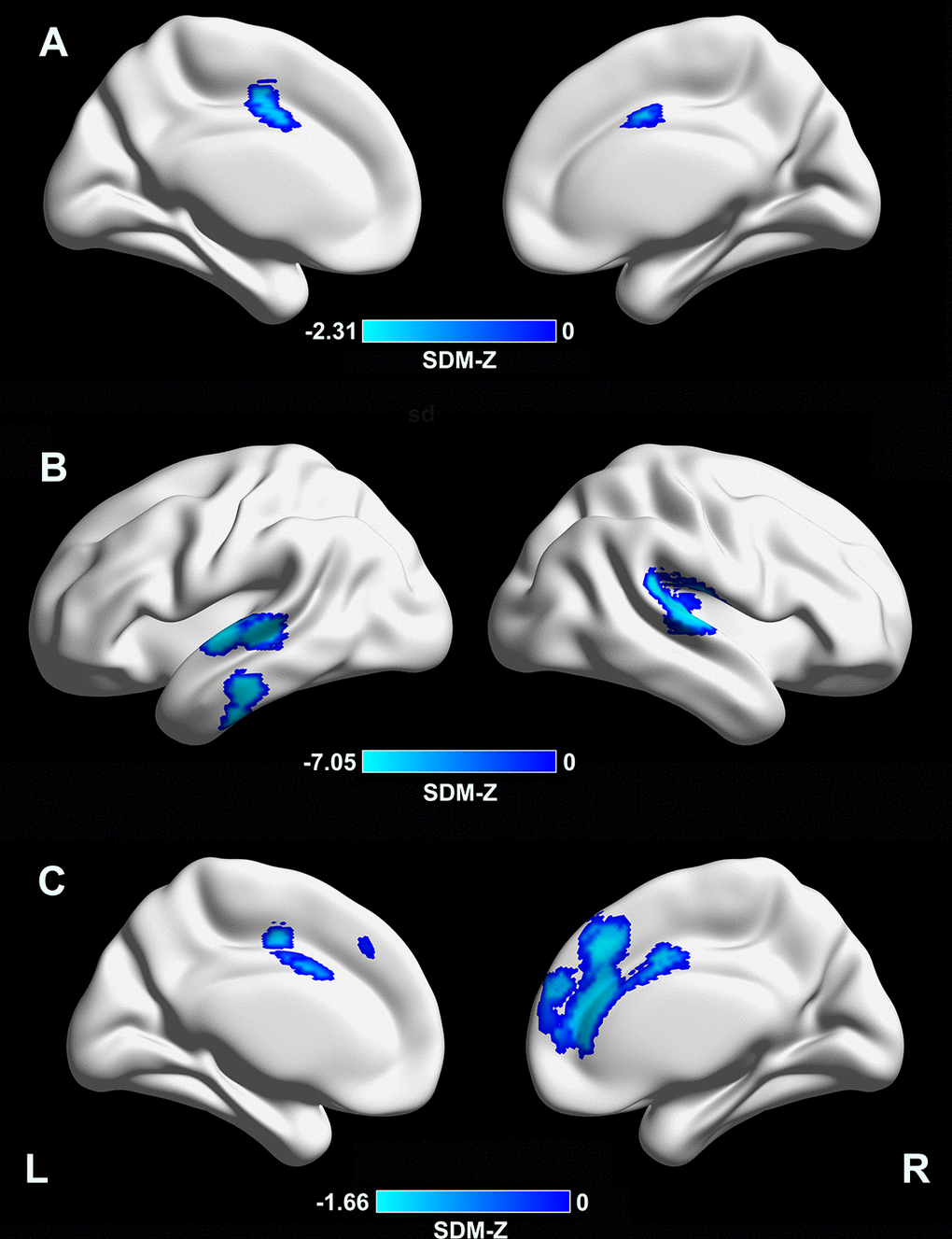
Figure 2.Meta-regression analyses of clinical variables with cortical thickness. (A) A longer disease duration was associated with lower CTh in the supplementary motor area/cingulate cortex (MNI coordinates: x = –4, y = –2, z = 46; BA 24; SDM-Z = –2.31; TFCE-based FWE corrected p = 0.009; voxels = 392). (B) A lower MMSE score in the PD sample was associated with lower CTh in the right superior temporal gyrus/rolandic operculum (MNI coordinates: x = 54, y = –22, z = 12; BAs 48 and 22; SDM-Z = 7.05; TFCE-based FWE corrected p = 0.001; voxels = 999), left superior/middle temporal gyri (MNI coordinates: x = –62, y = –12, z = 0; BAs 48, 21, and 22; SDM-Z = 6.65; TFCE-based FWE corrected p = 0.009; voxels = 441), and left inferior temporal gyrus (MNI coordinates: x = –60, y = –20, z = –24; BAs 20 and 21; SDM-Z = 6.57; TFCE-based FWE corrected p = 0.03; voxels = 169). (C) A higher LEDD in the PD sample was associated with lower CTh in the medial prefrontal cortex/anterior cingulate cortex (MNI coordinates: x = 4, y = 32, z = 38; BAs 32, 24, 10, and 8; SDM-Z = –1.66; TFCE-based FWE corrected p = 0.029; voxels = 1441). CTh, cortical thickness; MNI, Montreal Neurological Institute; BA, Brodmann area; SDM, seed-based d mapping; TFCE, threshold-free cluster enhancement; FWE, family-wise error; PD, Parkinson’s disease; LEDD, levodopa equivalent daily dose.