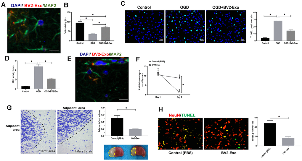
Figure 3.M2-phenotype microglia-derived exosomes (BV2-Exo) attenuated neuronal apoptosis induced by ischemic injury. (A) BV2-Exo was internalized by neurons in vitro, as imaged by confocal microscopy. Red color indicated exosomes (PKH26), blue color indicated nuclei (DAPI), and green color indicated neurons (MAP2). Scale bar = 25 μm. (B) Cell viability of neurons treated with OGD or OGD plus BV2-Exo, as determined by CCK-8 assay. (C) TUNEL assay for detecting apoptosis in neurons treated with OGD or OGD plus BV2-Exo. Scale bar = 20 μm. (D) Lactate dehydrogenase (LDH) assay for detecting LDH activity in neurons treated with OGD or OGD plus BV2-Exo. (E) BV2-Exo was internalized by neurons in vivo, as imaged by confocal microscopy. Red color indicated exosomes (PKH26), blue color indicated nuclei (DAPI), and green color indicated neurons (MAP2). Scale bar = 25 μm. (F) Modified neurological severity score for mice treated with control treatment (PBS) and BV2-Exo after tMCAO. (G) Relative infarct volume in brains of mice treated with control treatment (PBS) and BV2-Exo after tMCAO, displayed as brain cresyl violet staining and brain tissues of ischemic mice treated with indicated treatments. Yellow dotted boxes represent the infarct areas. (H) Double-staining of NeuN/TUNEL in brain sections of mice treated with control treatment (PBS) and BV2-Exo after tMCAO. Red color indicated NeuN and green color indicated TUNEL staining. Scale bar = 25 μm. Data are presented as mean±SD. *, p<0.05. At least three replicates were available for statistical analysis in each treatment.