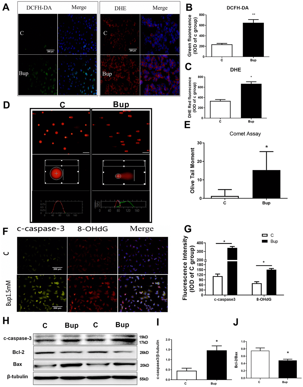
Figure 1.Bupivacaine induced SH-SY5Y cell oxidative DNA damage and neurotoxicity. After treating the SH-SY5Y cells with 1.5mM bupivacaine, the levels of intracellular reactive oxygen species (ROS) as stained by DCFH-DA (A, B) and superoxide anion as stained by DHE (A, C) were significantly increased. The index of DNA damage comet tail moment as assessed by in Comet Assay (D, E) were also robustly increased (* P <0.05). The oxidative DNA damage index 8-OHdG (F, G) increased. The expression of the apoptosis proteins cleaved-caspase3 (F, G, and H, I) increased while the apoptosis-related protein Bcl-2/Bax ratio (H, J) decreased. Data are the mean ± SD of three independent experiments, each performed in triplicates (*P <0.05, **P<0.01 vs control (C) group).