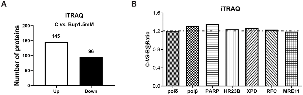
Figure 2.The DNA damage repair proteins expression of SH-SY5Y cells after exposure to bupivacaine were detected by iTRAQ proteomic screening. As showed in graph (A) iTRAQ proteomic screening results showed that: Of the total identified proteins, 241 proteins are significantly different between the (1.5 mM) bupivacaine and Control (C) groups, which included 145 upregulated and 96 downregulated proteins. Graph (B) displayed the list of DNA repair proteins whose expressions were increased by 1.2-fold or more after bupivacaine treatment (i.e., a ratio of Bup-vs-C greater than 1.2).