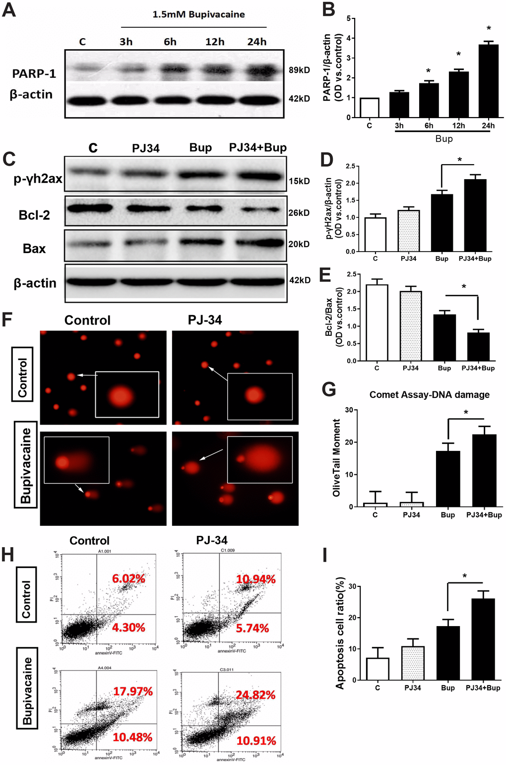
Figure 3.The key DNA repair protein PARP-1 closely participated in the repair of oxidative DNA damage in neurons caused by bupivacaine. In in vitro, the expressions of key repair protein PARP-1 in the BER pathway were significantly increased following bupivacaine-induced neuronal oxidative DNA damage. And, inhibition of PARP-1 expression with PJ34(a specific inhibitor of PARP) significantly aggravated the bupivacaine neurotoxicity. After SH-SY5Y cells were exposed to 1.5mM bupivacaine, the protein expression of PARP-1 (A, B) was increased obviously in a time-dependent manner. In the meantime, the DNA damage was aggravated: The DNA damage marker - phosphorylation level of γ-H2AX was significantly increased (C, D), while the comet assay indicator -the olive tail moment was significantly increased (F, G) in the Bupivacaine group as compared to Control group, which was concomitant with a significant reduction of the ratio of Bcl-2/Bax proteins (C, E) and increases of apoptosis as assessed by flow cytometry (H, I). Data are the mean ± SD of three independent experiments performed in triplicate, (*P <0.05, **P<0.01 vs C group).