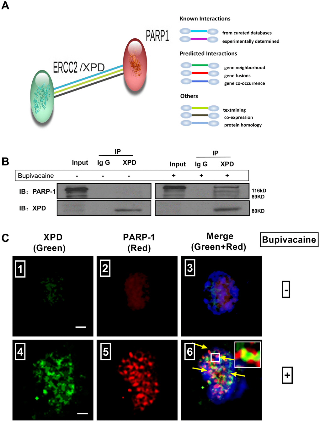
Figure 4.Possible existence of a novel interaction between XPD and PARP-1 in repairing the oxidative DNA damage caused by bupivacaine. Our previous study and iTRAQ results of the current study both suggested that XPD/ERCC2 also participated in the oxidative DNA damage of neurons caused by bupivacaine. But the interaction between PARP-1 and XPD is still unclear. Firstly, we identified the interaction between PARP-1 and XPD(ERCC2) using the STRING version 11.0 Program. (A) Combined screenshots came from the STRING website. Colored lines between the proteins indicate the various types of interaction evidence (according to the STRING website for color legend). Protein nodes that are enlarged indicate the availability of 3D protein structure information. No direct evidence or experimental data is available to confirm whether there exists an interaction between PARP-1 and ERCC2/XPD. For further verification, immunoprecipitation was applied to SH-SY5Y cells exposed to bupivacaine, and a strong interaction between XPD and PARP-1 was observed (B). Further, Immunofluorescence staining showed the colocalization (C ⑥, the yellow arrows) of XPD (Green) and PARP-1 (Red) following bupivacaine treatment in SH-SY5Y cells. Nuclei were stained with DAPI (blue). Bars, 5μm. Representative results of three experiments are shown.