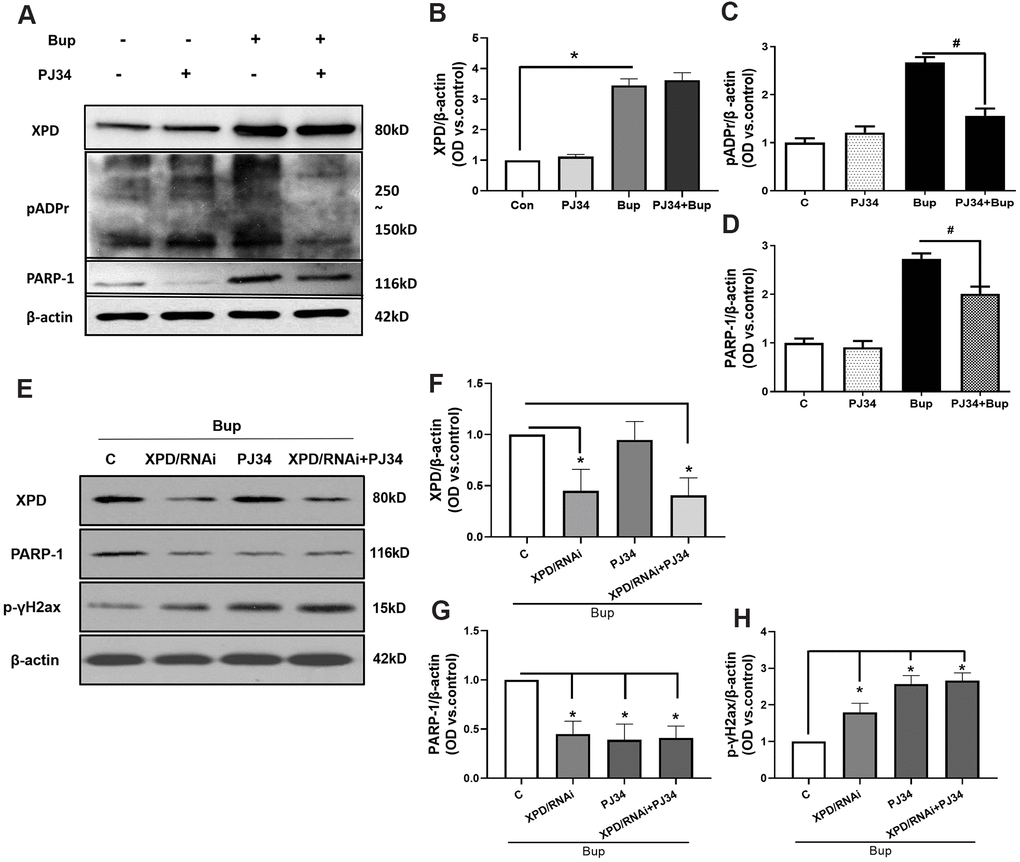
Figure 5.PARP-1 via XPD-mediated interaction contributed to the repairing of bupivacaine-induced neuron oxidative DNA damage. The protein expression of XPD and PARP-1 was examined in the presence or absence of PJ34(PARP-1 inhibition). Compared with the bupivacaine treatment alone group (Bup), the protein expression of pADPr (which represents the activation of PARP-1) was suppressed in cells treated with both PJ34 and Bup (PJ34+Bup group). After bupivacaine treatment, the expression of PARP-1 and XPD increased significantly. PJ34 can significantly reduce the expression and activity of PARP-1 caused by bupivacaine (A, C, D), but does not significantly inhibit the expression of XPD (A, B). Furthermore, the XPD-GV211-RNAi lentivirus was used to suppress the expression of XPD, while GV211-NC served as the control lentivirus group. PJ34 was used as the specific inhibitor of PARP-1. The expression of XPD was not affected after inhibition of PARP-1(E, F). However, after inhibiting the expression of XPD, PARP-1 expression was significantly reduced (E, G). Inhibition of either XPD or PARP-1 alone could increase the expression of the DNA damage index p-γ-H2AX induced by bupivacaine. However, concomitant inhibition of both XPD and PARP-1 did not further increase DNA damage (E, H). Data are the mean ± SD of three independent experiments performed in triplicate (*P <0.05, vs C group; #P<0.05, vs Bup group).