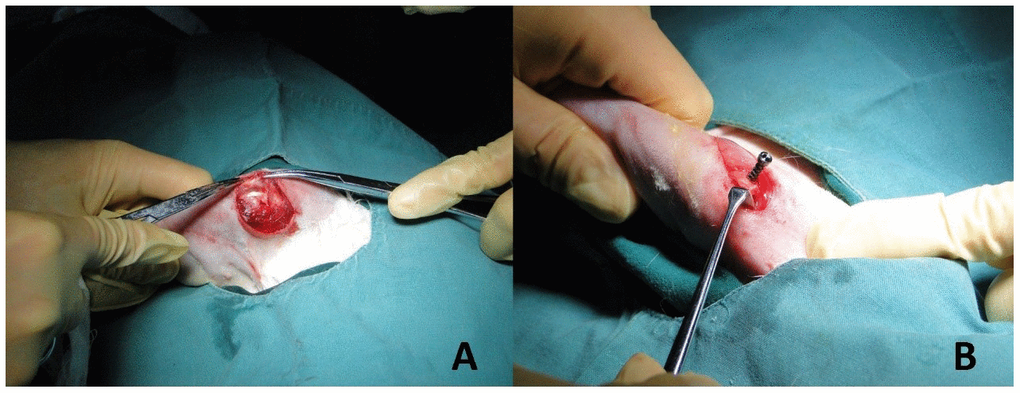
Figure 2.Establishment of implant model. (A) The deltoid muscle of the rabbit was bluntly dissected to reveal the greater tubercle of humerus. (B) Titanium alloy screws with different pore structures (trabecular pore structure in the study group and classical pore structure in the control group) were drilled in the long axis of the distal humerus approximately 120 degrees. The implant models of titanium alloy with different spatial structures of proximal humerus were established.