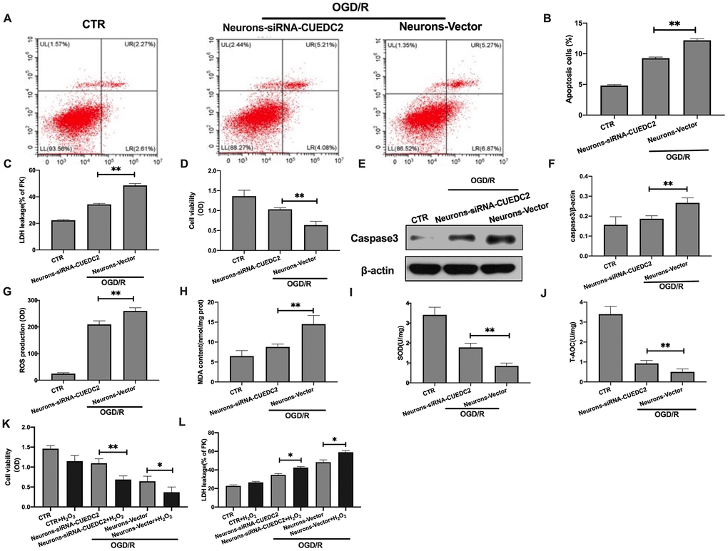
Figure 2.Effects of CUEDC2 on cerebral I/R-induced neuron insult. (A, B) Apoptotic cell death in neurons as detected by flow cytometry with Annexin V/PI staining. (C) Apoptotic cell death in neurons as detected by LDH-leakage assay. (D) Neuron viability as assessed by the MTT assay. (E, F) Protein expression of caspase-3 in neurons as assayed by western blotting. (G) ROS production in neurons as detected by DCFH-DA assay. (H) MDA production in neurons as evaluated by lipid peroxidation MDA assay. (I) SOD production in neurons as determined by WST-8 assay. (J) T-AOC level in neurons as detected by ABTS assay. (K) Viability of H2O2-treated neurons as analyzed by MTT assay. (L) Apoptotic cell death in H2O2-treated neurons as detected by LDH leakage assay. CTR, control; CUEDC2: CUE domain-containing 2; OGD/R: oxygen-glucose deprivation (4 hours) and reperfusion (24 hours); neurons-siRNA-CUEDC2: small interfering RNA silencing CUEDC2 in neurons; neurons-vector: the vector of neurons. All data are presented as the mean value ± SD. *p<0.05, **p<0.01; compared to the vector group, the H2O2 treatment group.