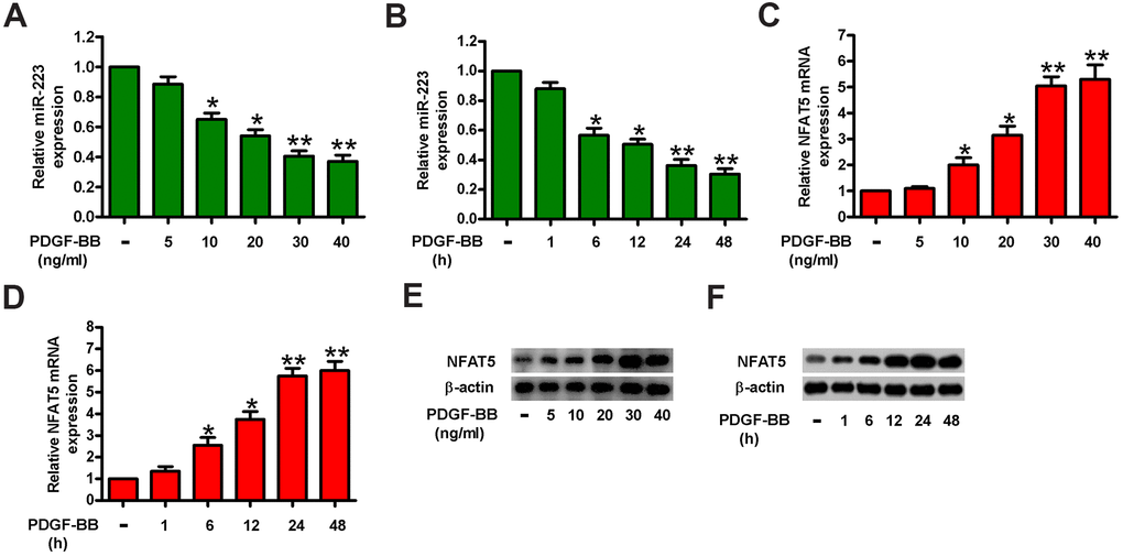
Figure 1.MiR-223 was increased and simultaneously NFAT5 was decreased in PDGF-BB-treated HASMCs. HASMCs were starved with 0.5% FBS for 48 h and subsequently exposed to PDGF-BB at various concentrations (5, 10, 20, 30, and 40 ng/ml) for the different time durations (1, 6, 12, 24, and 48 h). (A–D) miR-223 and NFAT5 mRNA levels were measured by qPCR assays in HASMCs treated with different concentrations of PDGF-BB (A, C) for the indicated time durations (B, D). MiR-223 and NFAT5 mRNA expressions were normalized to U6 and GAPDH, respectively. (E, F) Western blot analyses of NFAT5 expression in HASMCs treated with various doses of PDGF-BB (E) for the different time durations (F). β-actin was used as the endogenous control. The data are shown as mean ± SD of three separate experiments. *P < 0.05, **P < 0.01 compared with control group.