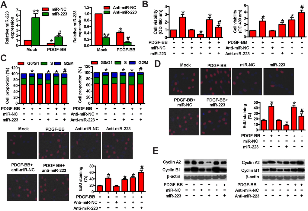
Figure 2.MiR-223 suppressed PDGF-BB-induced proliferation of HASMCs. Serum-deprived HASMCs were transfected with miR-223, miR-NC, and anti-miR-223 or anti-miR-NC for 24 h, followed by PDGF-BB stimulation for 24 h. (A) miR-223 levels were determined by qPCR assay. MiR-223 expression was normalized to U6. (B) MTT assay was performed to measure cell viability. (C) Cell cycle distribution was analyzed by flow cytometry. The percentage of cells in G0/G1, S, and G2/M phases were calculated. (D) Cell proliferation was assessed by EdU incorporation assay. The percentages of EdU-positive cells were counted. (E) Representative Western blot results of cyclin A2 and cyclin B1. β-actin was used as the endogenous control. The data are shown as mean ± SD of three separate experiments. *P < 0.05, **P < 0.01 compared with the miR-NC or anti-miR-NC group in (A) and compared with control group in (B–E). #P < 0.05 compared with PDGF-BB group.