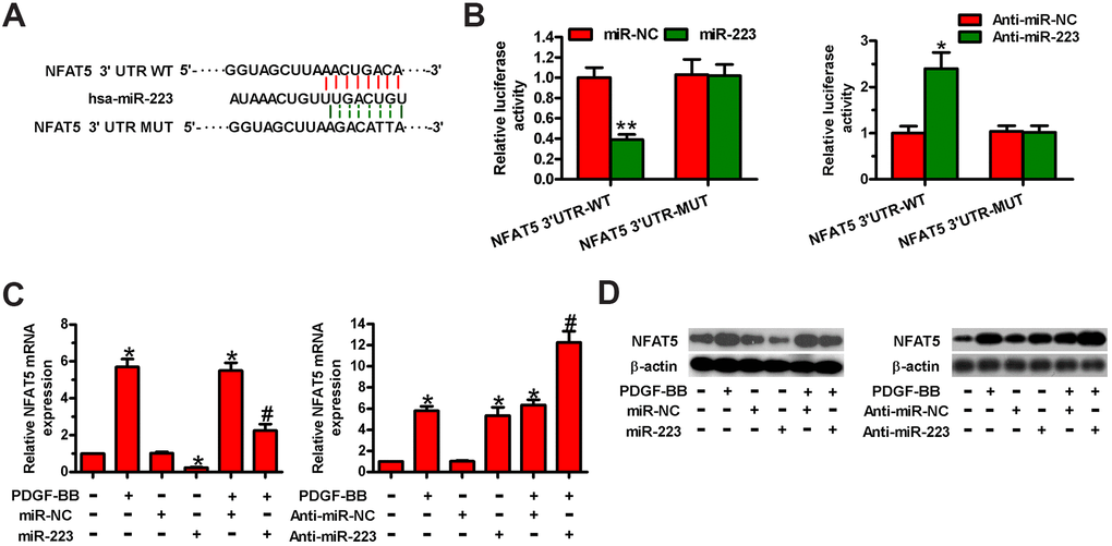
Figure 4.MiR-223 directly targeted NFAT5 in HASMCs. (A) The putative miR-223 binding sites in the 3′-UTR of NFAT5. (B–D) HASMCs were co-transfected with WT or MUT 3′-UTR of NFAT5 reporter plasmids or miR-223, anti-miR-223, miR-NC, or anti-miR-NC for 48 h. (B) Relative luciferase activity was detected. (C) qPCR and (D) Western blot assays were performed to assess NFAT5 mRNA and protein levels. GAPDH and β-actin were used as the endogenous controls, respectively. The data are shown as mean ± SD of three separate experiments. *P < 0.05, **P < 0.01 compared with the miR-NC or anti-miR-NC group in (B) and compared with control group in (C, D). #P < 0.05 compared with PDGF-BB group.