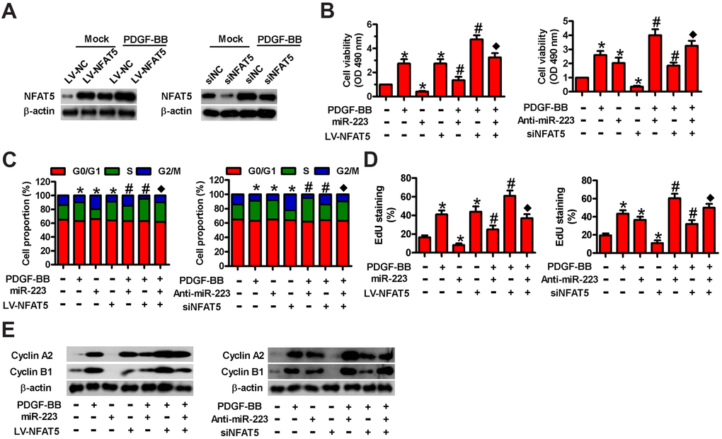
Figure 5.Involvement of NFAT5 in miR-223-elicited proliferative inhibition of PDGF-BB-exposed HASMCs. (A) Serum-deprived HASMCs were infected with LV-NC or LV-NFAT5 or transfected with siNC or siNFAT5 for 24 h, followed by PDGF-BB stimulation for 24 h. Western blot assay was conducted to detect NFAT5 expression. β-actin were used as the endogenous control. (B–E) Serum-deprived HASMCs were co-treated with miR-223 and LV-NFAT5 or anti-miR-223 and siNFAT5 for 24 h, followed by PDGF-BB stimulation for 24 h. (B) MTT, (C) flow cytometry and (D) EdU incorporation assays were carried out to analyze cell viability, cell cycle progression and proliferation. (E) Representative Western blot results of cyclin A2 and cyclin B1. β-actin was used as the endogenous control. The data are shown as mean ± SD of three separate experiments. *P < 0.05 compared with control group. #P < 0.05 compared with PDGF-BB group. ♦P < 0.05 compared with PDGF-BB + miR-223/anti-miR-223 or PDGF-BB + LV-NFAT5/siNFAT5 group.