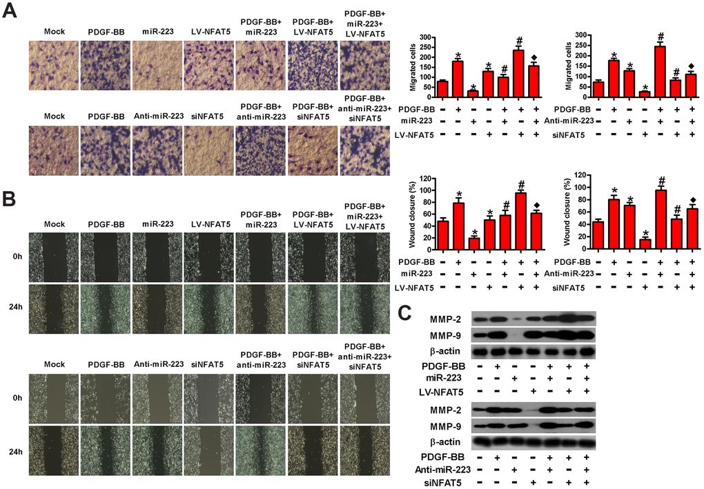
Figure 6.MiR-223 repressed the migration of PDGF-BB-exposed HASMCs by targeting NFAT5. Serum-deprived HASMCs were co-treated with miR-223 and LV-NFAT5 or anti-miR-223 and siNFAT5 for 24 h, followed by PDGF-BB stimulation for 24 h. (A) Transwell and (B) wound healing assays were conducted to measure cell migration. (C) The expression of MMP-2 and MMP-9 was detected by Western blot assays. β-actin was used as the endogenous control. The data are shown as mean ± SD of three separate experiments. *P < 0.05 compared with control group. #P < 0.05 compared with PDGF-BB group. ♦P < 0.05 compared with PDGF-BB + miR-223/anti-miR-223 or PDGF-BB + LV-NFAT5/siNFAT5 group.