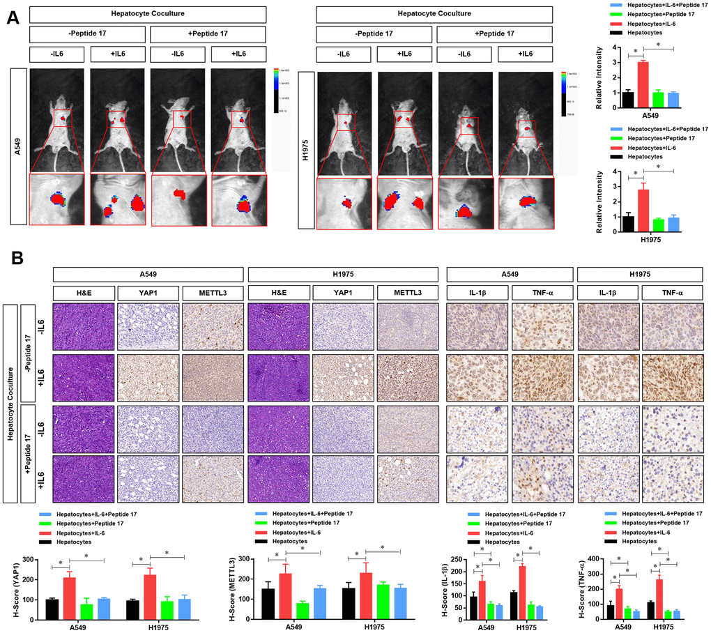
Figure 7.YAP1 blockade significantly inhibited the effects of the hepatic inflammatory microenvironment on the metastasis of lung adenocarcinoma (LUAD) cells. (A) Forty mice were divided into 8 groups, with 5 mice in each group. Cell grouping was similar to that of in vitro experiments, which were stimulated by the hepatic immune microenvironment and/or treated with the YAP1 inhibitor peptide 17 for 24 hours. After these treatments, 5×10⁶ cells each mouse was injected by tail vein and bioluminescence imaging (BLI) was performed 25 days later using the NightOWL II LB 983 imaging system (Berthold). (B) At the end of the experiment, all animals were sacrificed under anesthesia, and the metastasis tumors were fixated by Formalin and used for HE or IHC staining of YAP1 (×200), METTL3 (×200), IL-1b (×300) and TNF-a (×300). The quantitative analysis of IHC were performed by H-score scoring (*P<0.05 versus the control group).