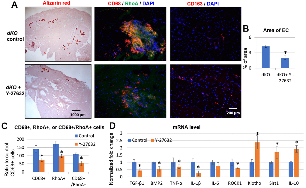
Figure 6.Systemic inhibition of RhoA/ROCK in dKO mice reduced calcification in skeletal muscle by repressing the accumulation of RhoA+/CD68+ cells. (A) Alizarin Red staining indicating reduction in calcification in dystrophic muscles of dKO mice treated with the RhoA/ROCK inhibitor Y-27632 for 3 times a week, from 3 weeks to 8 weeks of age. Immunostaining of skeletal muscle tissues showed that the accumulation of CD68+/RhoA+ cells in dKO muscle was also reduced by RhoA/ROCK inhibition, while the number of CD163+ cells (M2 macrophages) was increased. (B) quantification of the area of ectopic calcification (EC) in skeletal muscle of dKO mice with or without Y-27632 treatment. (C) Quantification of the number of CD68+, RhoA+, and CD68+/RhoA+ cells with and without Y-27632 treatment (number of cells per area of 100 myofibers). (D) qPCR results of mRNA isolated from dystrophic muscles of dKO mice showing that Y-27632 treatment significantly down-regulated the expression of pro-inflammatory/fibrosis genes (TGF-β1, BMP2, TNF-α, and IL-1β), and up-regulated the expression of anti-inflammatory/fibrosis genes (Klotho, Sirt1, and IL-10). n=8 for both dKO mice with or without Y-27632 treatment, * indicates p<0.05.