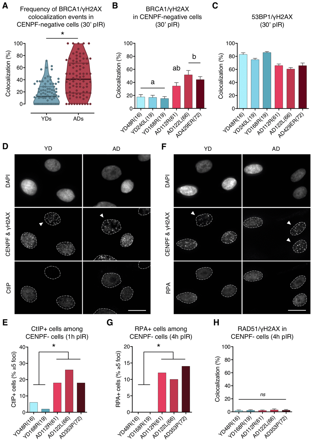
Figure 4.BRCA1, CtIP and RPA but no RAD51 are ectopically recruited to DNA DSBs in G1 cells from aged donors. (A) Percentage of BRCA1/γH2AX foci colocalization in CENPF-negative cells at 30 min after irradiation for each age group. Each dot corresponds to the fraction of BRCA1 and γH2AX foci colocalizing within one pore and the mean and quartiles are indicated (* p < .0001; n is stated in Supplementary Table 4; Mann–Whitney test). (B) Summary values for individual donors. Error bars indicate SEM (a≠b p < .05; n is stated in Supplementary Table 4; Kruskal–Wallis + Dunn). (C) Percentage of 53BP1/γH2AX foci colocalization for individual donors at 30 min after irradiation. Error bars indicate SEM (n is stated in Supplementary Table 2). (D) Immunofluorescent labeling of cell nuclei (DAPI), CENPF (A532), γH2AX (A594) and CtIP (A488). Arrowheads indicate G1 (CENPF-negative) cells. Scale bar = 20 μm. (E) Frequency of CtIP-positive HMECs (≥ 5 foci) at 1 h after irradiation (5 Gy, γ-rays). Analysis was restricted to CENPF-negative cells (* p < .05; n ≥ 50 cells/donor; Fisher’s exact test). (F) Immunofluorescent labeling of cell nuclei (DAPI), CENPF (A532), γH2AX (A594) and RPA (A488). Arrowheads indicate G1 (CENPF-negative) cells. Scale bar = 20 μm. (G) Frequency of RPA-positive HMECs (≥ 5 foci) at 4 h after irradiation (5 Gy, γ-rays). Analysis was restricted to CENPF-negative cells (* p < .05; n ≥ 50 cells/donor; Fisher’s exact test). (H) Percentage of RAD51/γH2AX foci colocalization in CENPF-negative cells at 4 h after irradiation (5 Gy, γ-rays). Error bars indicate SEM (nsp > .05; n ≥ 1000 γH2AX foci/donor; Kruskal–Wallis + Dunn).
Figure 4 — Age-associated deficient recruitment of 53BP1 in G1 cells directs DNA double-strand break repair to BRCA1/CtIP-mediated DNA-end resection | Aging