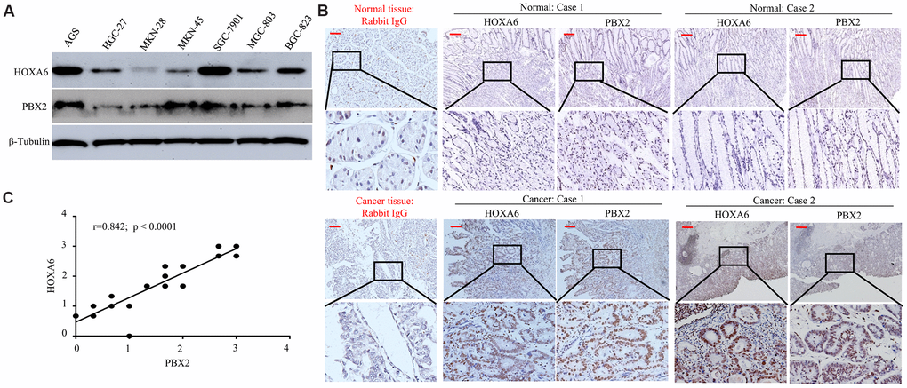
Figure 6.The positive expression of HOXA6 correlated with PBX2 expression. (A) Western blot analysis was is used to determinate the expression of HOXA6 and PBX2 in GC cell lines. (B) Representative IHC images of HOXA6 and PBX2 in GC (n =23) and their matched adjacent tissues following serial sectioning. The first antibody used for normal rabbit IgG as an isotype control. (C) Spearman correlation analysis of HOXA6 and PBX2 protein levels in GC tissue. Scale bars in B represent 50 μm.