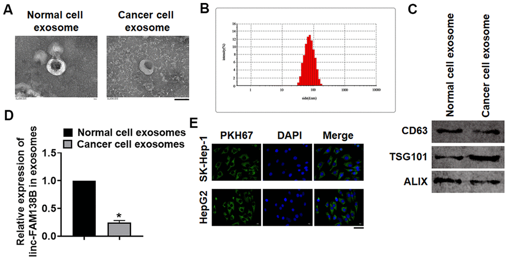
Figure 2.Linc-FAM138B was packaged into exosomes from cancer cells in HCC. Normal and cancer cells were isolated from HCC tissues, then exosomes were isolated from the supernatant of normal and cancer cells. (A) TEM images of exosomes isolated from normal and cancer cells. Scale bar, 100 nm. (B) Zetasizer Nano ZS was used to detect the diameter of isolated exosomes. (C) Western blot for exosome markers CD63, Tsg101 and Alix. (D) The expression of linc-FAM138B in exosomes from normal cells and cancer cells was tested by qRT-PCR. (E) PKH67 labeled linc-FAM138 was transfected into tumor cells. Then, SK-Hep-1 and HepG2 were incubated with exosomes from supernatant of tumor cells. And immunofluorescence experiment indicated a dominant fluorescence intensity of PKH67 in SK-Hep-1 and HepG2 cells. Scale bar, 100 μm. Data are mean ± SD; *P < 0.05. Data among multiple groups were analyzed by one-way ANOVA, followed by a Tukey post hoc test. The experiment was repeated in triplicate.