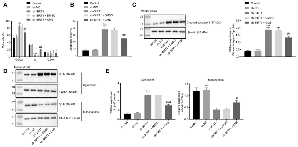
Figure 7.CMS suppressed the apoptosis of HG-treated hRMECs. hRMECs were treated with sh-NC, sh-SIRT1, sh-SIRT1 + DMSO and sh-SIRT1 + CMS, respectively. (A) Cell cycle distribution as determined by flow cytometry analysis. (B) Cell apoptosis as determined by flow cytometry analysis. (C) Representative Western blots of cleaved caspase-3 protein and its quantitation in hRMECs, normalized to β-actin. (D, E) Western blots of Cyt-C protein (D) and its quantitation (E) in the cytoplasm and mitochondria, normalized to β-actin. *p < 0.05, **p < 0.01, ***p < 0.001, compared to sh-NC-treated cells, and #p < 0.05, ##p < 0.01, ###p < 0.001, compared to cells stimulated with sh-SIRT1 + DMSO. The results were the measurement data and expressed as mean ± standard deviation. Comparisons between multiple groups should be analyzed by one-way ANOVA with Tukey's post hoc test. The cell experiments were repeated three times independently. CMS, coumestrol; HG, high glucose; hRMECs, human retinal microvascular endothelial cells; NC, negative control; DMSO, dimethyl sulfoxide; SIRT1, sirtuin 1; NO, nitric oxide; Cyt-C, cytochrome c; ANOVA, analysis of variance.