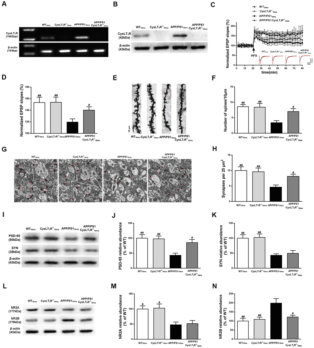
Figure 2.CysLT1R deficiency enhances hippocampal synaptic plasticity in APP/PS1 mice. (A) Western blot detection of CysLT1R protein in mice hippocampus. (B) RT-PCR assay of CysLT1R mRNA in mice hippocampus. (C) The induction of hippocampal LTP was assessed after high-frequency stimulation (HFS; indicated as an arrow) and recorded for 60 min post-induction. (D) Summary bar-graphs showing differences in mean values of fEPSPs slope during 55-60 min following the induction of LTP among genotypes. (E) Representative images of Golgi-impregnated dendrites in the hippocampus. Scale bar = 10 μm. (F) Statistical analysis of the average number of dendritic spines. (G) The synaptic density in the hippocampus was determined by electron microscopy. Scale bar = 1 μm. (H) Statistical analysis of synaptic density calculated as the number of synapses per 25 μm2. (I) Representative immunoblots of PSD-95 and SYN in mice hippocampus. Quantifications of (J) PSD-95 and (K) SYN protein levels were expressed as the ratio (in %) of WT group. (L) Representative immunoblots of NR2A and NR2B in mice hippocampus. Quantifications of (M) NR2A and (N) NR2B were expressed as the ratio (in %) of WT group. All values are expressed as mean ± SEM, n = 4-6, #P<0.05, ##P<0.01, ###P<0.001 vs. APP/PS1 mice.