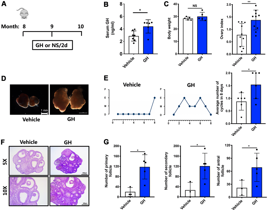
Figure 3.Effects of GH administration in vivo on the ovarian reserve. (A) Schematic illustration for the NS and GH-treated mice. (B) The GH levels in the peripheral blood was measured in GH (n = 6) and vehicle (n = 7) group. (C) The changes of body weight and ovary index after GH administration. NS, no significance. (D) Micrographs of NS-treated and GH-treated mouse ovaries. Scale bar, 1 mm. (E) Left: Estrous cycle in representative females. Right: Average numbers of cycles in 8 days in two groups. P, proestrus; E, estrus; M, metestrus; D, diestrus. (F) HE-stained of NS-treated and GH-treated mouse ovaries. Scale bar, 400 μm, 200 μm. (G) Follicle counts and the number of corpus luteum in NS-treated (n = 3) and GH-treated (n = 5) mice. Data are presented as mean ± SD. *P < 0.05, **P < 0.01.