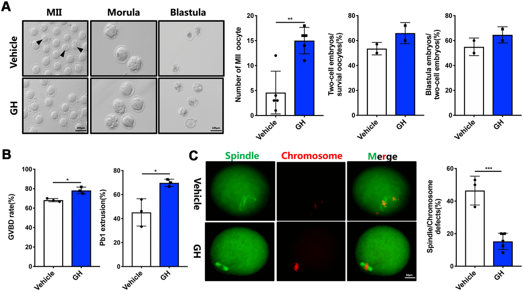
Figure 4.Effects of GH treatment in vivo on the quality and meiotic progress of aged oocytes. (A) Left: Representative images of MII oocytes, morula and blastocysts from NS-treated and GH-treated mice. Black arrows point to fragmented MII oocytes. Scale bar, 200 μm, 100 μm. Right: Number of MII oocytes and percentage of 2-cell embryos and blastocysts in NS-treated and GH-treated mice. (B) After cultured in M2 medium, the rate of GVBD and Pb1 extrusion were recorded. (C) Left: The MII oocytes from NS-treated (n = 37) and GH-treated (n = 42) mice were stained with α-tubulin (green) and propidium iodide (PI) (red). Scale bar, 50 μm. Right: Quantification of NS-treated and GH-treated oocytes with abnormal spindle/chromosomes. Data are presented as mean ± SD. *P < 0.05, **P < 0.01, ***P < 0.001.