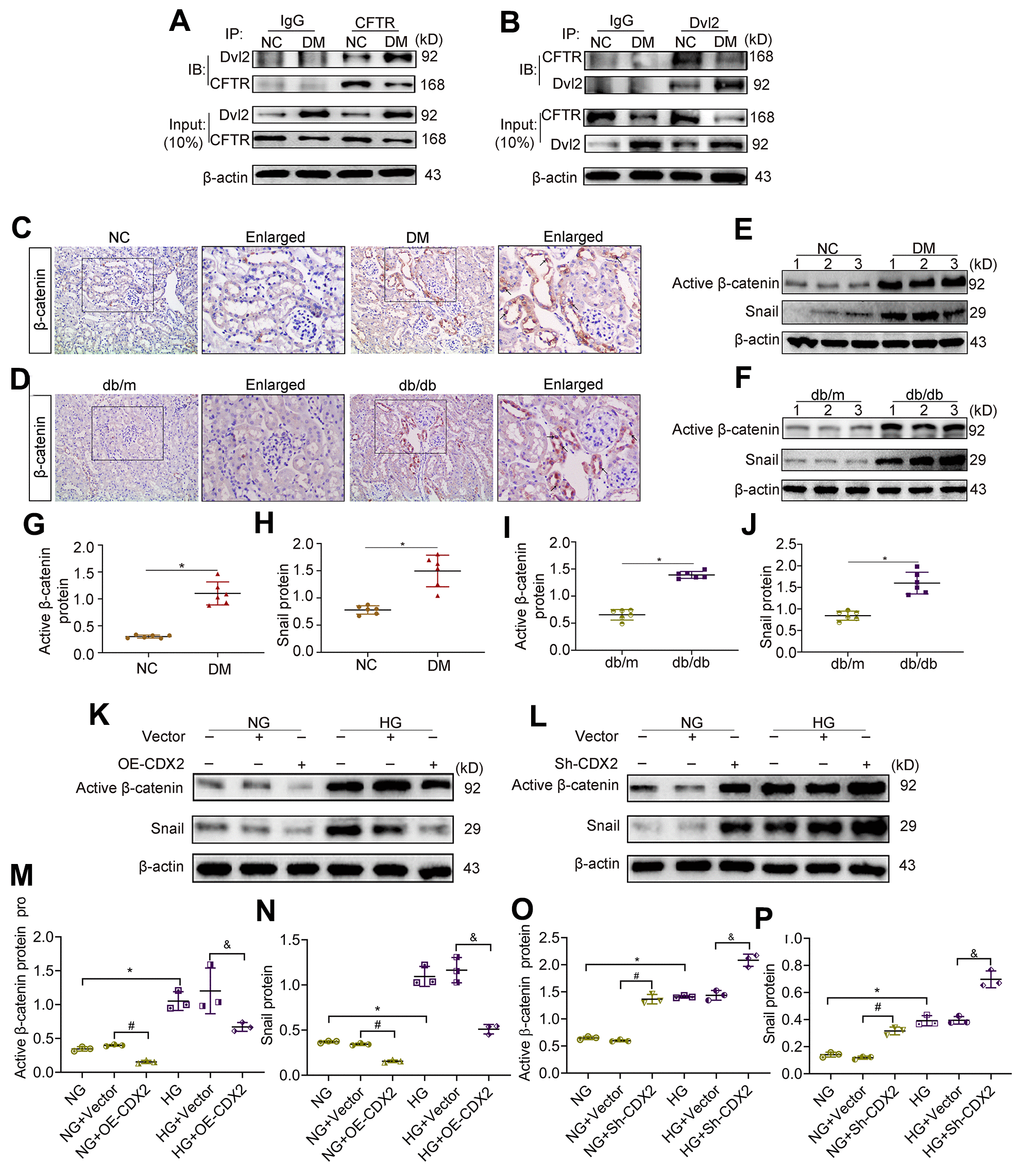
Figure 5.Downregulation of CFTR activates β-catenin and causes renal fibrosis. (A, B) Co-immunoprecipitation assay indicated that CFTR and Dvl2 interacted with each other in vivo. The Input group was a positive control group. In the CO-IP group, the kidney tissue lysates of T1D mice and controls were immunoprecipitated with IgG, anti-CFTR (A) or anti-Dvl2 (B) antibodies, and the resulting immunoprecipitates were blotted (IB) with anti-CFTR and anti-Dvl2 antibodies. The protein samples used for co-immunoprecipitation were normalized to β-actin. (C, D) Immunohistochemical staining of β-catenin in T1D model mice and controls (C), and T2D mice and controls (D). Positive staining (black arrow) (magnification, ×200); enlarged box area (magnification, ×400). (E–J) Western blot bands of Activated β-catenin and Snail in T1D model mice and controls (E), and T2D model mice and controls (F); quantitative data are shown (G–J). n=6; *P<0.05 versus NC group or db/m group. (K–P) Western blot bands (K, L) and quantitative data (M–P) of activated β-catenin and Snail in non-transfected (NG group, HG group) NRK-52E cells, and NRK-52E cells transfected with Vector (NG+Vector group or HG+Vector group), or CDX2-overexpressing (NG+OE-CDX2 group or HG+OE-CDX2 group) or CDX2-knockdown (NG+Sh-CDX2 group or HG+Sh-CDX2 group) plasmid. Data are mean±SD from three experiments performed independently. n=3; *P<0.05 versus NG group; #P<0.05 versus NG+Vector group; &P<0.05 versus HG+Vector group.