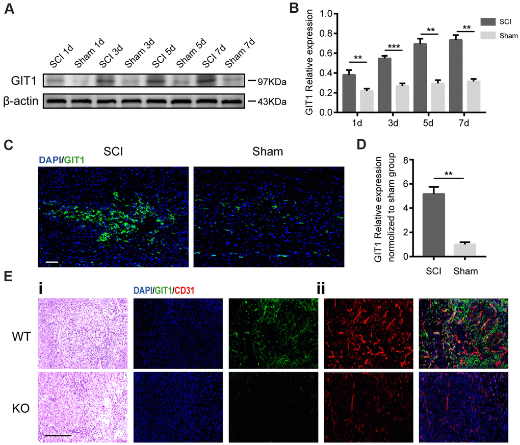
Figure 1.GIT1 is highly expressed in vivo after SCI. (A, B) Western blots and semiquantification of GIT1 at each time point after SCI or sham surgery. (C, D) Immunofluorescence images for GIT1 (green) and quantification at day 7 after SCI or sham surgery. Nuclei were counterstained using DAPI (blue). Scale bar, 100 μm. (E) Spinal cord at 7 days post-SCI: (i) representative H&E images in the lesion site from the GIT1 WT and KO groups. Bar, 100 μm. (ii) IF staining of GIT1 (green) and CD31 (red) in the lesion epicenter from the GIT1 WT and KO groups. Nuclei were stained using DAPI (blue). Scale bar, 100 μm. N = 5 animals per group. ***p < 0.01, ***p < 0.001.