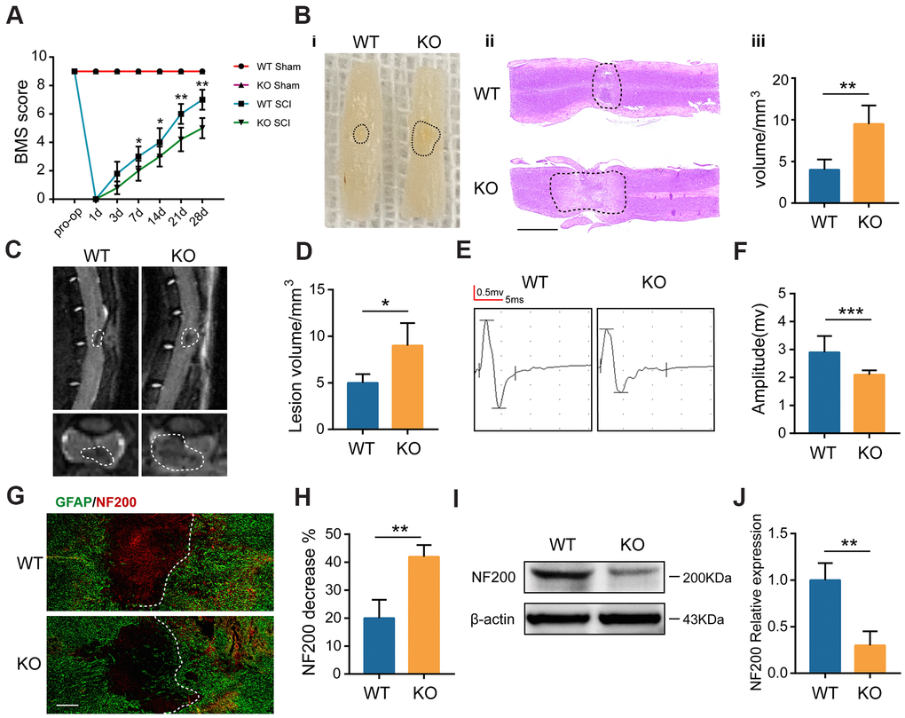
Figure 2.GIT1 deficiency exacerbates SCI-induced spinal cord damage. (A) Basso Mouse Scale results over 28 days. (B) Spinal cord at 28 days post-SCI: (i) gross morphology, (ii) representative H&E stained sections. Scale bar = 2000 μm, and (iii) the lesion volumes. (C, D) Sagittal and axial spinal cord T2 weighted images at day 28 after SCI. (E, F) MEP analysis. (G, H) IF staining of GFAP (green) and NF200 (red) at the injury sites at 7 days after SCI. Nuclei were stained using DAPI (blue); the dashed lines represent the boundary of the injury area. Scale bar = 100 μm. (I, J) Representative western blots of NF200 and the semiquantification of relative expression levels of NF200. N = 6 animals in each group. *p < 0.05, **p < 0.01, ***p < 0.001.