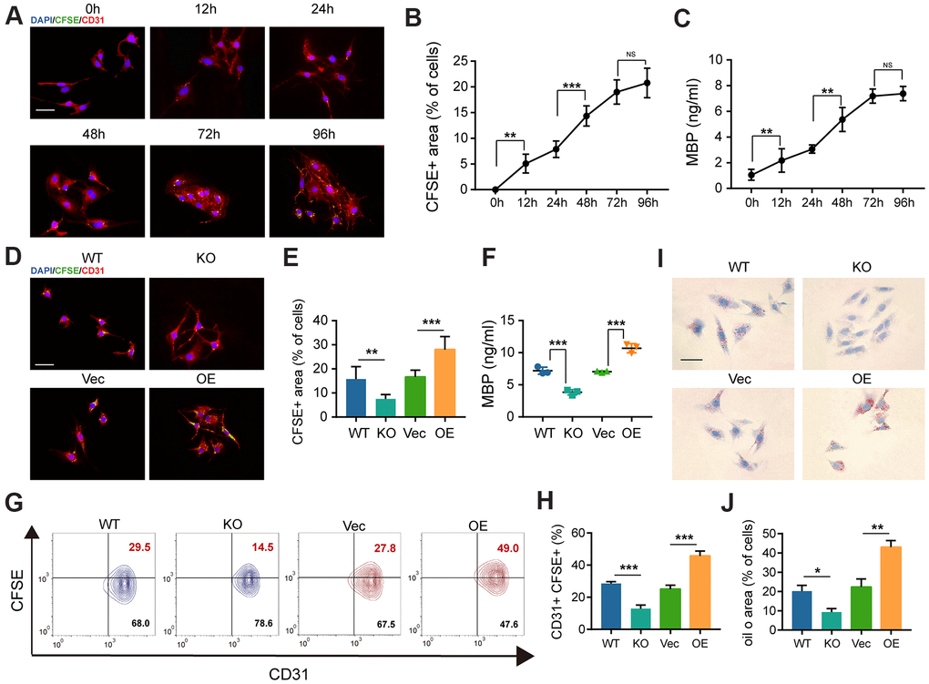
Figure 5.The deficiency of GIT1 in BMECs fails to engulf and degrade myelin debris efficiently in vitro. (A) Representative IF images showing CFSE-labeled myelin debris (green) engulfed by BMECs (red) at indicated time points. Nuclei were stained using DAPI (blue). Scale bar = 20 μm. (B) Phagocytosis of myelin debris (the percentage of the CFSE positive area). (C) ELISA results of intracellular MBP after being cultured with or without myelin debris at indicated time points. (D, E) IF staining and quantification of phagocytosis of CFSE-labeled myelin debris by BMECs at 72 h. Scale bar = 20 μm. (F) ELISA detection of intracellular MBP in different BMECs after being cultured with myelin debris for 72 h. (G, H) FACS detection of myelin-laden BMECs at 72 h. The CD31+ and CFSE+ quadrant represents myelin-laden BMECs. (I) Oil Red O staining of BMECs in the different groups after being cultured with myelin debris for 72 h. Scale bar = 20 μm. (J) Quantification of the ORO stained positive areas of BMECs from the different groups after being stimulated with myelin debris for 72 h. N = 6 in each group. NS represents no significance, *p < 0.05, **p < 0.01, ***p < 0.001.