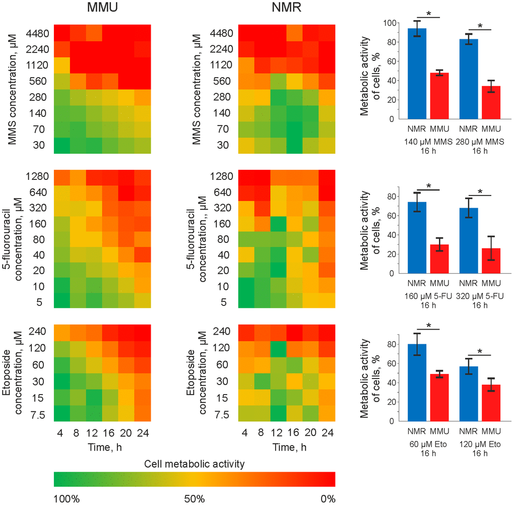
Figure 1.Metabolic activity of mouse and NMR fibroblasts upon treatment with 5FU, MMS and Eto. Mouse (MMU) and NMR fibroblasts, NSF8 and 3T3 cells, respectively, were treated with indicated DNA damaging agents. The conditions of treatment (time and concentrations of cytotoxic agents) are indicated on the plot. The color changing from green to red indicates the decrease in metabolic activity. The metabolic activity of untreated cells was taken as 100% and corresponds to the green color on the plot. Right side: a major decrease in metabolic activity, which was about 20-40%, is shown for the treatment of NMR fibroblasts with 280 μM of MMS, 320 μM of 5FU and 120 μM of Eto. These results are presented together with the decrease of metabolic activity in mouse fibroblasts upon treatment with the same concentrations of DNA damaging agents. Abbreviations: Mouse (MMU); 5-fluorouracil (5FU), etoposide (Eto), and methyl methanesulfonate (MMS). The standard deviation is shown; the confidence is based on the Mann-Whitney U test, *P < 0.05.