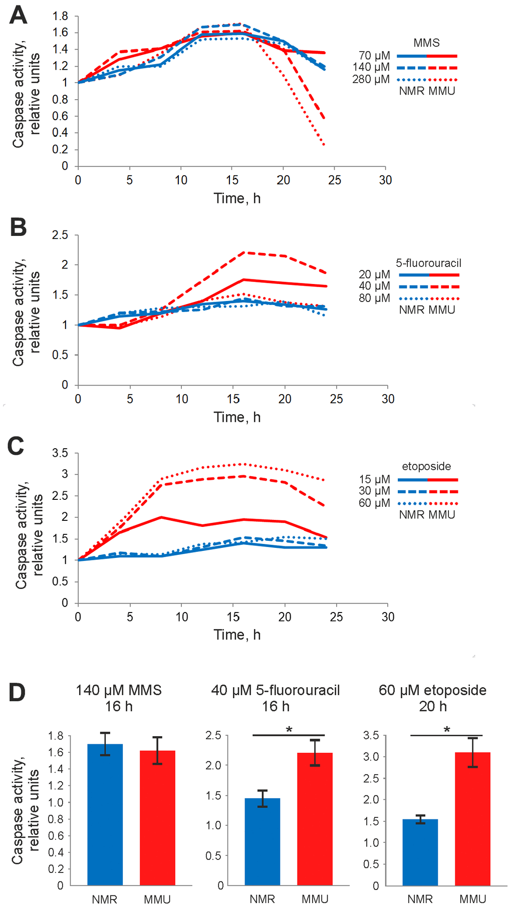
Figure 2.Caspase activity of NMR and mouse fibroblasts upon treatment with DNA damaging agents determined by Caspase-Glo 3/7 assay. Activity of effector caspases-3/7 was measured after treatment with: (A) 70 μM (solid line), 140 μM (dashed line), and 280 μM (dotted line) MMS; (B) 20 μM (solid line), 40 μM (dashed line) and 80 μM (dotted line) 5FU; (C) 15 μM (solid line), 30 μM (dashed line), and 60 μM (dotted line) Eto. The caspase activity in untreated cells was taken as one relative unit. Results represent the mean of at least three independent experiments. (D) Caspase activity of NMR and mouse cells is shown for treatment with 140 μM MMS for 16 h, 40 μM 5FU for 20 h and 60 μM Eto for 16 h. Standard deviation is shown; confidence is based on the Mann-Whitney U test, *P < 0.05. The activity of NMR and mouse cells is shown in blue and red, respectively. Abbreviations: Mouse (MMU); 5-fluorouracil (5FU), etoposide (Eto), and methyl methanesulfonate (MMS).