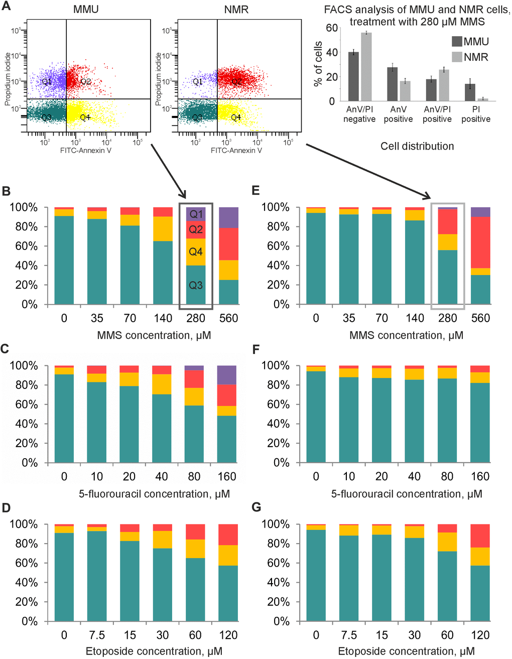
Figure 3.Flow cytometry analysis of MMU and NMR cells upon treatment with DNA damaging agents. (A). NMR and mouse cells were stained with FITC-Annexin V (AnnV) and Propidium Iodide (PI). The gating strategy for double negative AnnexinV-/PI-(Q3), single positive, early apoptotic cells (AnnexinV+/PI-) (Q4), single positive (AnnexinV-/PI+) (Q1) and double positive AnnexinV+/PI+ (Q2) is shown. (B–G). Cell death of MMU (B–D) and NMR (E–G) cells under the wide spectrum of cytotoxic conditions was evaluated by FACS analysis. The total number of cells was taken as 100%; blue color indicates the population of viable cells, double negative AnnexinV-/PI-(Q3); yellow color is used for single positive, early apoptotic cells (AnnexinV+/PI-) cells (Q4); red color is implemented for double positive AnnexinV+/PI+ cells (Q2); purple color for single positive (AnnexinV-/PI+) cells (Q1). The representative experiment out of three independent ones is shown.