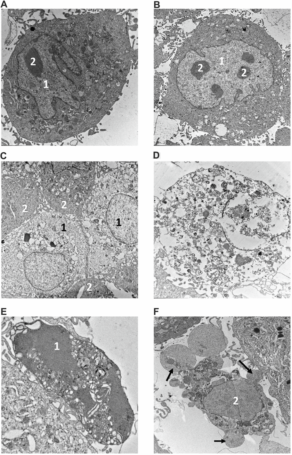
Figure 4.Electron microscopy analysis of MMU and NMR cells upon treatment with MMS. (A, B). The images of cells from NMR and mouse, that remained untreated, are shown. 1 – nucleus, 2 – nucleolus. (C–F). The images of cells after incubation with MMS. (C) swollen NMR cells (1) with cytoplasm of low electron density; and cells without signs of alteration (2) are shown. (D) necrotic mouse cell is shown, in which organelles are completely destroyed. Apoptosis of NMR (E) and mouse (F) cells. NMR cell nucleus (1) with condensed chromatin and organelles is shown, mouse cell demonstrates extensive “apoptotic blebbing” (“apoptotic blebs” are shown with arrows), 2 – nucleus without chromatin condensation but altered morphology.