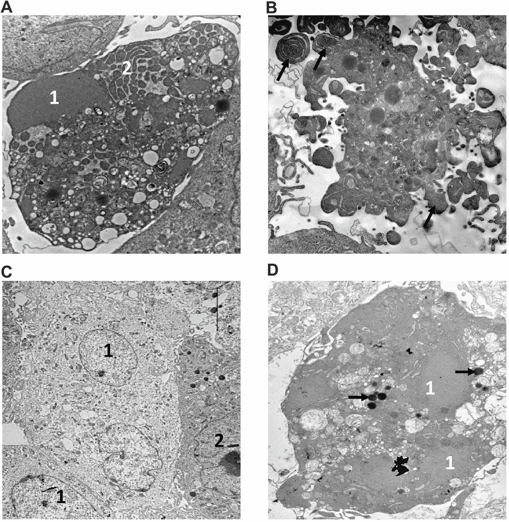
Figure 5.Electron microscopy analysis of MMU and NMR cells upon treatment with 5FU and Eto. Apoptotic features of cells from NMR (A) and mouse (B) incubated with 160 μM of 5FU are shown. The NMR cell is filled with the remnants of the nucleus (1) with condensed chromatin and organelles; the endoplasmic reticulum area is marked with the number "2". (B) section of mouse cell periphery demonstrates extensive apoptotic blebbing” (“apoptotic blebs” are shown with arrows). (C, D). The images of NMR cells treated with Eto are shown. (C) swollen cells (1) with cytoplasm of low electron density, and cell without signs of alteration (2). (D) apoptotic cell with a highly condensed nucleus (1) and cytoplasm. Organelles are not seen; arrows show lipid droplets.