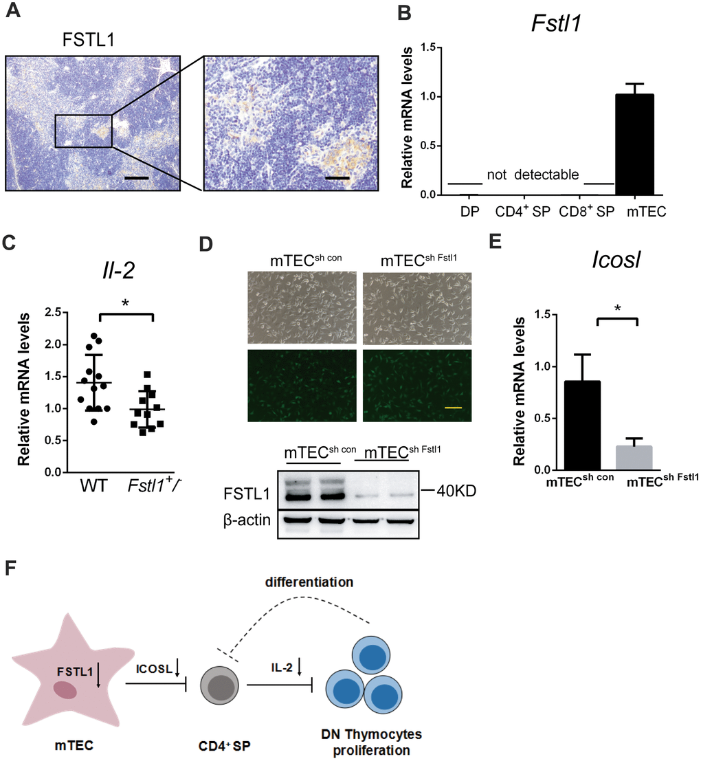
Figure 5.Deficiency of FSTL1 in mTEC cells inhibited the production of IL-2 by CD4+ SP. (A) Representative micrographs of FSTL1 IHC staining of thymus slices from 8-week-old WT mice. Scale bar, 200 μm (left), 50 μm (right). (B) Results of qRT-PCR showing mRNA level of Fstl1 in DP, CD4+ SP, CD8+ SP thymocytes and mTEC cells. (C) Results of qRT-PCR showing mRNA levels of Il-2 in WT and Fstl1+/- mouse thymuses, The gene mRNA level was normalized to that of β-actin. (D) mTEC cells were infected with lentiviral vectors encoding Fstl1 specific shRNAs (sh Fstl1) or control vector (sh CON). The infection was indicated by green fluorescent protein (upper), and the infection efficiency was evaluated using western blot (lower). (E) Results of qRT-PCR showing mRNA level of Icosl in mTECsh con groups and mTECsh Fstl1 groups. The gene mRNA level was normalized to that of β-actin. (F) Schematic illustration of the proposed mechanism of action of deficiency of FSTL1 on mTEC cells to decrease the proliferation of DN thymocytes and impair the development of T cells. Knockdown of Fstl1 in mTEC cells inhibited the expression of Icosl, which directly interacted with CD4+ SP thymocytes to decrease the production of IL-2, inhibiting DN thymocyte proliferation. Further, the decreased proliferation of DN thymocytes might inhibit the differentiation into CD4+ SP thymocytes. Data are presented as mean ± SD. Each dot in the graphs represents an individual mouse. Data in the bar chart represents three sets of independent experiments. *p < 0.05.