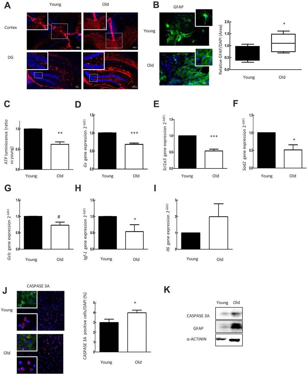
Figure 1.Aged astrocytes have increased reactivity, inflammation and cell death as well as loss of nutritive and anti-oxidative capacity. (A) Representative immunofluorescence for GFAP (red) in cortex and DG of young (2 month-old) and aged (over 24 month-old) C57BL/6J mice (n=2). (B) Representative immunofluorescence and the quantification for GFAP positive cells in 1DIV (young) and 30DIV (old) primary astrocytes cell culture derived from neonatal Wistar (n=6). (C) ATP luminescence levels of young and old primary astrocytes cultures (ratio compared to the young group) (n=4). (D–I) Expression of gs, slc1a3, sod2, gclc, igf-1 and il-6 in young and old primary astrocytes cultures (n=6). (J) Representative immunofluorescence of CASPASE 3A and co-staining of CASPASE 3A (red) with GFAP (green) together with DAPI (blue). Quantification for CASPASE 3A positive cells and in 1DIV (young) and 30DIV (old) primary astrocytes cell culture derived from P1 Wistar rat pups (n=6). (K) Protein expression of CASPASE 3A and GFAP in 1DIV (young) and 30DIV (old) primary astrocytes cell culture. Results are expressed as the mean ± SEM. Asterisks denote the significance levels when compared to the control group (***p<0.001, **p<0.01 and *p<0.05 versus controls, t-test).