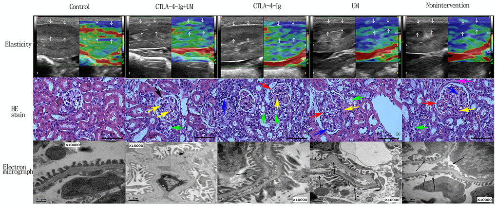
Figure 2.Elastic imaging, H&E staining, and electron micrograph analyses of right kidney parenchymal podocytes in rats. Assessment of rat renal parenchymal elasticity. Control group: The majority of the region of interest (arrow) is green, with a small portion being red; score = 1. CTLA-4-Ig + UM group: The majority of the region of interest (arrow) is green, with some areas being red and blue; score = 2. CTLA-4-Ig group: The majority of the region of interest (arrow) is green, with some areas being red and blue; score = 2. UM group: The region of interest (arrow) is primarily blue with some green; score = 3. Non-intervention group: The region of interest (arrow) is primarily blue with some green; score = 3. H&E staining of renal parenchymal tissue samples. -Control group: No glomerular capillary cavity changes, cellular proliferation, or basement membrane thickening are evident, with clear glomerular balloon; -CTLA-4-Ig + UM group: Glomerular volume is slightly enlarged and the glomerular basement membrane is partially thickened (black arrow), with slight cellular proliferation, with a small amount of hyaline substance deposition (yellow arrow) and vacuolated degeneration of the renal tubular epithelial cells (green arrow); CTLA-4-Ig group: Glomerular basement membrane thickening is evident (red arrow), with cellular proliferation, a small amount of hyaline substance deposition (yellow arrow), narrowing of the partial capillary lumen, marked vacuolated degeneration of the renal tubular epithelial cells (green arrow), and interstitial lymphocyte infiltration (blue arrow); UM group: Glomerular volume enlargement and basement membrane thickening are evident (red arrow), with marked cellular proliferation, flaky hyaline substance deposition (yellow arrow), vacuolated degeneration of renal tubular epithelial cells (green arrow), and narrowing of the capillary lumen (blue arrow); Non-intervention group: Glomerular volume enlargement and basement membrane thickening are evident (red arrow), with cellular proliferation, flaky hyaline substance deposition (yellow arrow), vacuolated degeneration of the renal tubular epithelial cells (green arrow), narrowing of the capillary lumen (blue arrow), and hyalinosis of the glomerular wall (pink arrow). Assessment of podocyte ultrastructural features via TEM. (The splicing is used to join together two parts of the same TEM image due to the limitation of the field of view). Control group: Podocytes exhibit a uniform arrangement without any fusion or loss; CTLA-4-Ig + UM group: Podocyte synapses appear disorderly, without obvious fusion or loss; CTLA-4-Ig group: Podocyte synapse structures are still present, but with visible evidence of fusion (black arrow); UM group: Disorder of the podocyte synapse is evident, with some missing synapses, slight protrusion of the basement membrane, and visible synaptic fusion; Non-intervention group: the volume of the podocyte synapse is larger, with some missing and fused podocytes.