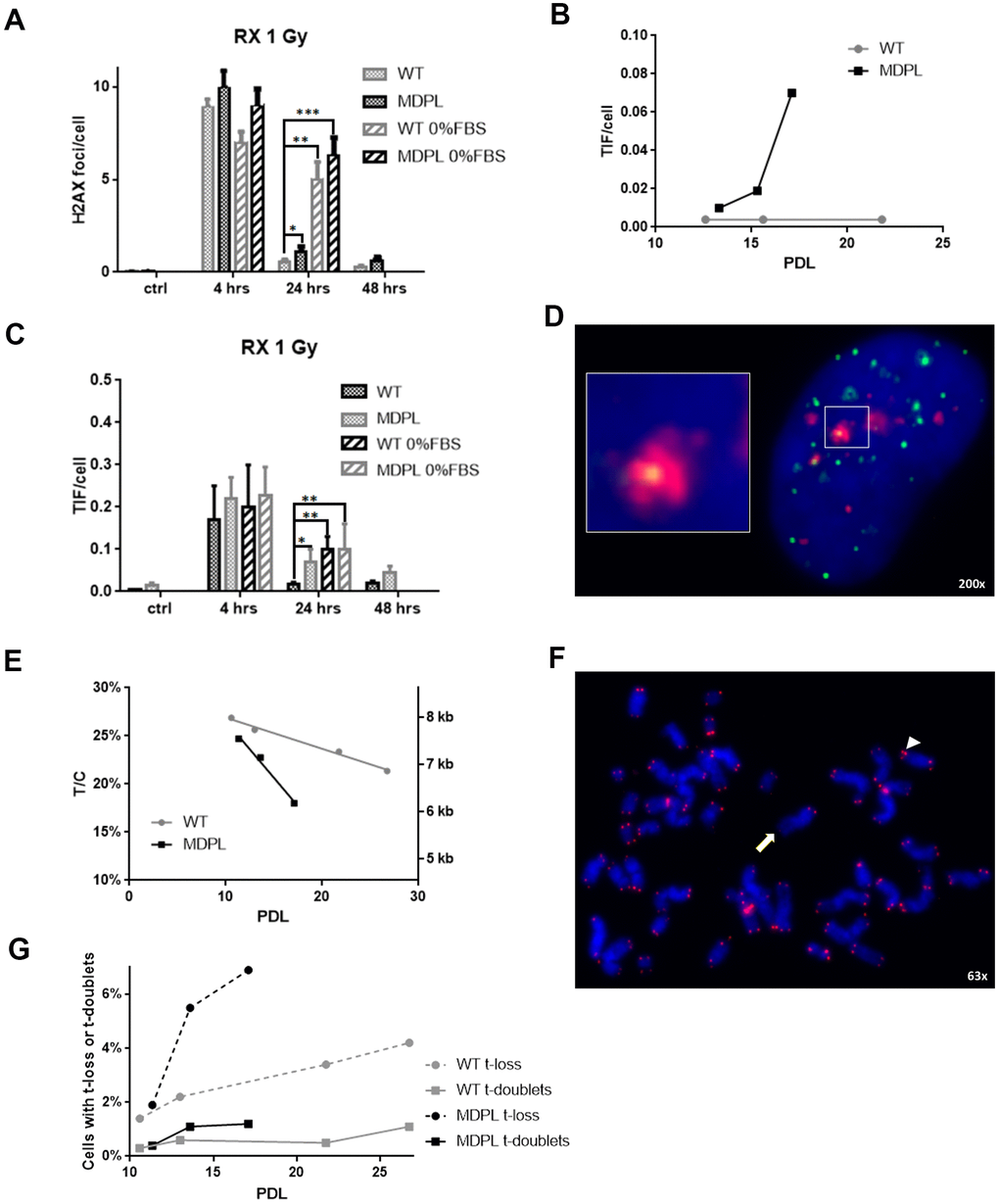
Figure 6.DNA repair kinetics after 1 Gy of X-irradiation. (A) DNA repair kinetics evidenced by γH2AX foci in serum-fed and serum-depleted cells after 1 Gy of X-irradiation. (B) Telomere-induced foci (TIF) in unirradiated fibroblasts at different population doubling levels (PDL). (C) Time course of TIF in serum-fed and serum-depleted cells after 1 Gy of X-irradiation. (D) Representative image of Telomeres stained with anti-TRF1 antibody (green), foci stained with anti-γH2AX antibody (red), and DAPI-stained nucleus (blue). Magnification 200X; inset shows co-localization of both antibodies, indicating a TIF. (E) Ratio between telomeric and centromeric fluorescence: T/C. (F) Representative image of chromosome spread showing telomere doublet (arrowhead) and telomere loss (arrow). Magnification 63X. (G) Telomere loss (circles, dashed lines) and telomere doublets (squares, continuous lines) at different PDL. WT in grey, MDPL in black.