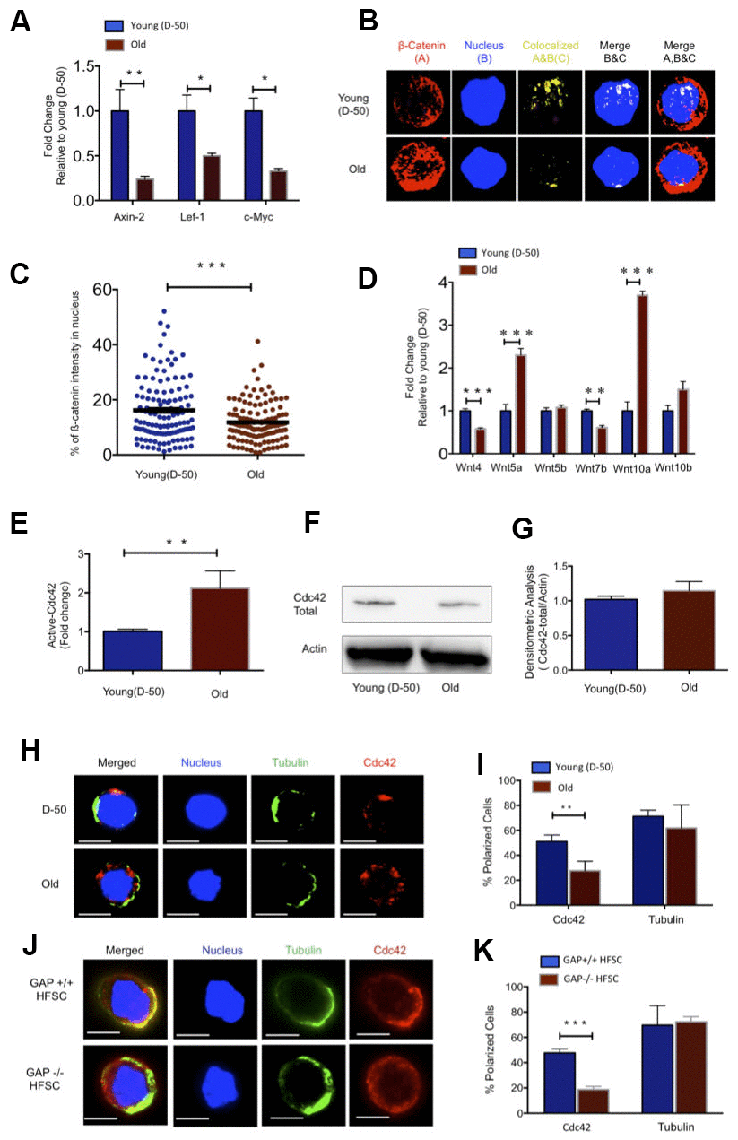
Figure 1.Increased Wnt5a expression in aged HFSC results in a shift from canonical to non-canonical Wnt signaling. (A) Expression of target genes of the canonical Wnt pathway in FACS sorted young and old HFSC (Sca-1-/lowA-6highCD34+), N≥3 (B) Z-stacks and three-dimensional merged images of ß-catenin (red), nucleus (dapi, blue) and co-localization signal (yellow) in FACS sorted young and old HFSC by Immunofluorescence (C) Quantification of the ß-catenin signal in the nucleus of young and old HFSC, N=4 (D) Wnt ligand transcript levels in young (D-50) or old (>2years) HFSC, N≥3 (E) ICdc42-activity levels in old Sca-1-/low keratinocyte lysate compared to young Sca-1-/low keratinocyte measured by G-LISA, N=4; (F) Cdc42 protein levels in young and old Sca-1-/low keratinocytes (representative Western blot) (G) Densitometry score of Cdc42 protein levels from Western blots, N=4 (H) Representative picture of Cdc42 (red) and tubulin (green) in young and old HFSC, scale bar = 5μm (I) Percentage of cells with a polar distribution of Cdc42 and tubulin in young and old HFSC, N≥6 (J) Representative picture of a non-polar distribution of Cdc42 (red) in young HFSC from Cdc42GAP-/- as compared to HFSC from young wild type Cdc42GAP+/+ mice, scale bar=5μm (K) Quantification of polar distribution (percentage) of Cdc42 and tubulin in HFSC from Cdc42GAP+/+ and Cdc42GAP-/- mice, N≥3, P<0.05, **P<0.01, ***P<0.001 (paired student’s t test). Error bars represent s.e.m.