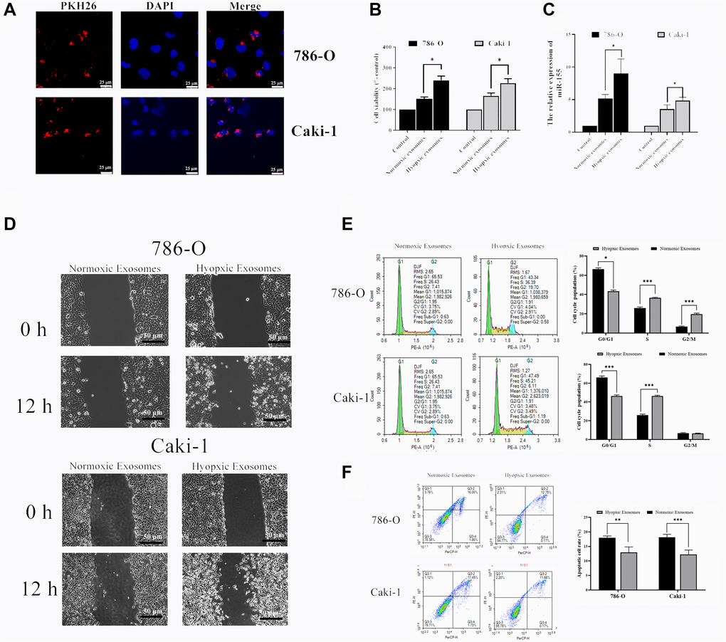
Figure 4.Hypoxia-induced RCC cell-derived EVs alter the viability of 786-O and Caki-1 cells. (A) EV uptake by 786-O and Caki-1 cells after 12 h was detected using PKH26-labeled EVs (red) and DAPI (blue) for nuclear localization. (B) The impact of hypoxia- and normoxia-derived EVs on the viability of RCC cells. The results indicated that hypoxia-induced EVs to significantly bolster the viability of treated RCC cells. (C) The impact of hypoxia- and normoxia-derived EVs on miR-155 levels. (D) Representative micrographs of the wound healing assay at 0 h and 12 h. Hypoxia-associated EVs derived from both of these cell lines were able to promote enhanced RCC cell migration. (E) Cell-cycle distribution was assessed by flow cytometry. Hypoxia-associated EV treatment was associated with an increased number of cells in S phase. (F) Apoptosis was assessed by flow cytometry. Hypoxia-associated EV treatment inhibited the cell apoptosis. This experiment was conducted using three distinct biological replicates. *P < 0.05, **P < 0.01, and ***P < 0.001.