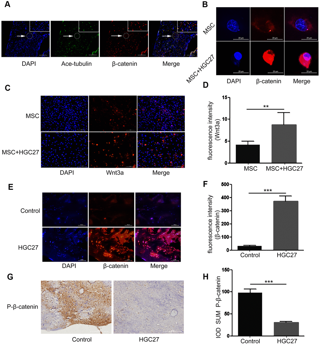
Figure 5.Gastric cancer activates the canonical Wnt/β-catenin signaling pathway in bone. (A) Immunofluorescence analysis of acetylated α-tubulin (ciliary axonin, green) and β-catenin (red) in femur from PBS injected C57BL mice. DAPI (nuclear marker, blue) staining was used as counterstain. Scale bars, 100 μm. (B) Immunofluorescence staining of MSCs with or without co-cultured HGC27 to visualize β-catenin (red) and nuclei (DAPI, blue) states. Scale bar, 20 μm. (C) Immunofluorescence staining of MSCs with or without co-cultured HGC27 showing Wnt3a (red) and nuclei (blue). Scale bar, 100 μm. (D) Quantification of immunofluorescence intensity in (C). (E) Representative femoral tissues from C57BL mice injected with PBS or HGC27 were analyzed for β-catenin expression by immunofluorescence staining at day 90. Scale bar, 50 μm. (F) Quantification of immunofluorescence intensity in (E). (G) Immunohistochemical staining for phosphorylated-β-catenin (p-β-catenin) in femurs of PBS or HGC27-injected groups. Scale bar, 100 μm. (H) IOD SUM of positive cells from (G) were compared between the HGC27 group and control femurs. Data are shown as mean±SEM. Statistical differences were obtained using Student's t-test, **, p<0.01, ***, p<0.001. n=3 per-group.