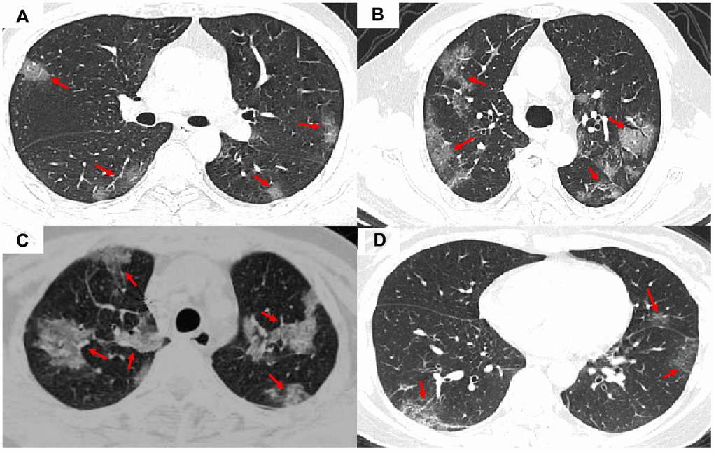
Figure 1.Representative images of COVID-19 pneumonia, adenovirus pneumonia, cytomegalovirus pneumonia, and influenza virus pneumonia. (A) A transverse CT image from a 35-year-old man with adenovirus pneumonia showing bilateral ground-glass opacities in the upper lobes with a rounded morphology (arrows). (B) COVID-19: A transverse CT image from a 57-year-old man with COVID-19 showing more limited ground-glass opacities in the bilateral upper lobes with an elliptical morphology (arrows). (C) A transverse CT image obtained in a 45-year-old female with cytomegalovirus pneumonia showing bilateral ground-glass and burr-like, denser, and less transparent distribution (arrows). (D) A transverse CT image of a 61-year-old man diagnosed with influenza virus pneumonia showing bilateral ground-glass opacities in the upper lobes (arrows).